
|
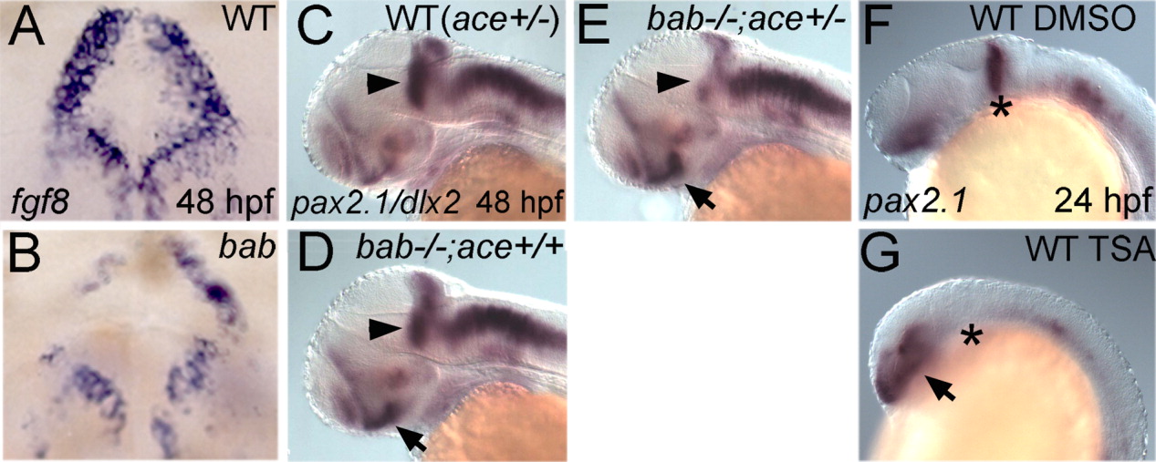
Fig. 6 rerea and fgf8 genetically interact in the midbrain-hindbrain boundary (MHB), and disruption of REREa or inhibition of histone deacetylase (HDAC) activity results in the inability to maintain the MHB. A-F: All panels show whole-mount in situ hybridizations in frontal view (A,B) or lateral view (C-G) of 48 hours postfertilization (hpf; A-E) or 24 hpf (F,G) embryos. Genotypes are shown in the upper right corner, whereas probes and stages are shown in the lower left and right corners, respectively. A,B: bab (B) displays degeneration of the MHB as seen by the patchy disappearance of fgf8-expressing cells compared with WT embryos (A). C,D: pax2.1 is expressed in the MHB (arrowheads) in wild-type (WT; ace+/-, C) and bab-/-;ace+/+ (D) embryos at 48 hpf. E: Reduction of Fgf8 in bab-/-;ace+/- leads to a reduction of pax2.1 in the MHB. dlx2 expression was used for the purpose of identifying bab embryos and does not interfere with the MHB staining of pax2.1. F,G: The 350 nM Trichostatin A (TSA) treatment from dome stage to 24 hpf also leads to a loss of pax2.1 expression at the MHB (asterisk in G) compared with wild-type embryos treated with dimethylsulfoxide (DMSO) alone (F). C-G: pax2.1 expression in the eye (arrows) is up-regulated in bab (D,E) and TSA-treated embryos (G) compared with wild-types (C,F).