
|
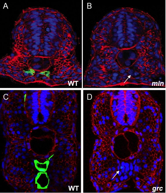
Fig. S1 The number of endothelial cells is reduced in mins202 and grcs635. (A–D) Transverse sections of 30 hpf Tg(flk1:EGFP)s843 wild-type (A) and mins202 mutant (B) embryos, and 36 hpf wild-type (C) and grcs635 mutant (D) embryos, visualized for Tg(flk1:EGFP)s843 expression (green), β-Catenin (red), and TOPRO (blue). White arrows point to the apparent lack of endothelial cells in mutant embryos (as assessed by Tg(flk1:EGFP)s843 expression). In wild-type embryos, 6 to 7 endothelial cells are present per any given optical section. In comparison, mins202 mutant embryos have less than one endothelial cell per section (n = 10), and grcs635 mutant embryos have less than 3 endothelial cells per section (n = 14).
Reprinted from Developmental Biology, 307(1), Jin, S.W., Herzog, W., Santoro, M.M., Mitchell, T.S., Frantsve, J., Jungblut, B., Beis, D., Scott, I.C., D'Amico, L.A., Ober, E.A., Verkade, H., Field, H.A., Chi, N.C., Wehman, A.M., Baier, H., and Stainier, D.Y., A transgene-assisted genetic screen identifies essential regulators of vascular development in vertebrate embryos, 29-42, Copyright (2007) with permission from Elsevier. Full text @ Dev. Biol.