
|
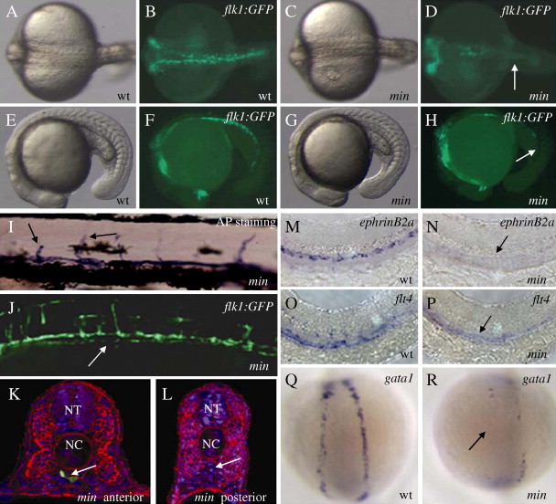
Fig. 2 mirinay regulates endothelial and hematopoietic lineages. (A–H) Bright-field (A, C, E, and G) and epifluorescent (B, D, F, and H) micrographs of 20 hpf wild-type (A, B, E, and F) and mins202 mutant (C, D, G, and H) embryos, shown in dorsal (A to D) and lateral (E to H) views. Note the reduction of endothelial cells in the posterior region of mins202 mutant embryos (arrows). (I–J) Micrographs of 96 hpf mins202 mutant larvae stained for endogenous alkaline phosphatase activity (I, bright-field) and visualized for Tg(flk1:EGFP)s843 expression (J). Black arrows point to arrested intersegmental vessels (SEs) (I), white arrow points to discontinuous axial vessel (J). (K, L) Transverse sections from anterior (K) and posterior (L) trunk of 36 hpf mins202 mutant embryos, visualized for Tg(flk1:EGFP)s843 expression (green), β-Catenin (red), and TOPRO (blue). Although the vasculature of mins202 mutant embryos eventually recovers, a drastically reduced number of endothelial cells is observed at this stage (white arrows point to the region of axial vessels). (M–R) Defective endothelial cell specification in 24 hpf mins202 mutant embryos (N and P) compared to wild-type embryos (M and O), as assessed by in situ hybridization with the arterial endothelial marker ephrinB2a (M and N), and the venous endothelial marker flt4 (O and P); and defective erythropoiesis in 18 hpf mins202 mutant embryos (R) compared to wild-type (Q), as assessed by examining gata1 expression in dorsal views. Black arrows in panels N and P point to the reduction in endothelial marker expression in mins202 mutant embryos, and black arrow in panel R points to the region of the lateral plate mesoderm where erythrocytes form in wild-type embryos. Abbreviations: NT: neural tube, NC: notochord.
Reprinted from Developmental Biology, 307(1), Jin, S.W., Herzog, W., Santoro, M.M., Mitchell, T.S., Frantsve, J., Jungblut, B., Beis, D., Scott, I.C., D'Amico, L.A., Ober, E.A., Verkade, H., Field, H.A., Chi, N.C., Wehman, A.M., Baier, H., and Stainier, D.Y., A transgene-assisted genetic screen identifies essential regulators of vascular development in vertebrate embryos, 29-42, Copyright (2007) with permission from Elsevier. Full text @ Dev. Biol.