
|
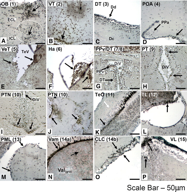
Fig. 3 Discrete clusters of pCREB+ cells localize to all proliferative zones of the adult zebrafish brain. Within the olfactory bulb [PZ1; OB; A], strongest immunoreactivity is observed within the external cellular layer (ECL), close to the junction with the telencephalon (white arrow). Scattered positive cells are also widespread within the inner cellular layer (ICL) and glomerular cell layer (not shown). Isolated pCREB+ cells are present within the ventral telencephalon [PZ2; VT; B], with the strongest immunoreactivity towards the base of the telencephalon, near the junction of the OB. A tighter demarcation of pCREB+ cells within the dorsal telencephalon [PZ3; DT; C] exists, localized exclusively to the dorsal region (Dd), with very few pCREB+ cells within the telencephalon proper (Dc). Two discrete clusters of pCREB+ cells are visible within the ventral region of the pre-optic area (POA), including the anterior parvocellular pre-optic nuclei (arrows) [PZ4; POA; D]. Within the ventral thalamus [PZ5; VeT; E], two discrete clusters of cells, lining the walls of the telencephalic ventricle (TeV), are positive for pCREB, whereas in the more dorsally located habenula [PZ6; Ha; F], three discrete clusters of pCREB+ cells are visible (arrows). Four proliferation zones are present lining the walls of the diencephalic ventricles (DiV) the periventricular pretectal nuclei [PZ7; PPv; G], dorsal thalamus [PZ8; DT; G], posterior tuberculum [PZ9; PT; H]; and the posterior tuberculum, located within the hypothalamus [PZ10; PTN; I (coronal)–J (sagittal)]. Within the optic tectum [PZ11; TeO; K] a tight band of pCREB+ cells is visible within the periventricular grey zone (PGZ), layers 1 and 2 (black arrow), whereas most cells within the TeO layer 3 are negative for pCREB (white arrow). Within the torus longitudinalis [PZ12; TL; L], most cells are pCREB+ (black arrow), excluding a discrete area of the ventral TL, although the ventral-most TL cells are all strongly positive for pCREB. All cells of the posterior mesencephalic lamina [PZ13; PML; M] are positive for pCREB, as are cells within the molecular layer of the valvula cerebelli [PZ14a; Vam; N]. Additionally, pCREB+ cells reside in the granular layer (white arrow), and in the cerebellar caudal lobe [PZ14b; CLC; O]. Within the vagal lobe [PZ15; VL; P], a cluster of pCREB+ cells is visible in the dorsal-most region (black arrow). Planes of section: sagittal (A–F, J–P) coronal (G–I). Scale bar 50 μm.
Reprinted from Developmental Biology, 307(1), Dworkin, S., Heath, J.K., Dejong-Curtain, T.A., Hogan, B.M., Lieschke, G.J., Malaterre, J., Ramsay, R.G., and Mantamadiotis, T., CREB activity modulates neural cell proliferation, midbrain-hindbrain organization and patterning in zebrafish, 127-141, Copyright (2007) with permission from Elsevier. Full text @ Dev. Biol.