
|
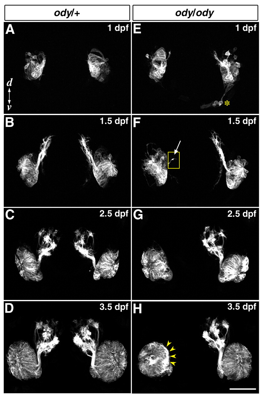
Fig. 9 Live imaging of axon projection in zebrafish ody mutant embryos. Developmental processes of axon projection in representative ody/+;omp:yfp/+ (A-D) and ody/ody;omp:yfp/+ (E-H) embryos are shown in frontal views. Yellow-boxed inset in F is shown at high gain to reveal faint axonal fibers. In the ody/ody embryo, pioneer axons extending toward the presumptive OB (arrow in F) from one of two olfactory placodes are significantly reduced in number at 1.5 dpf, when compared with those from the contralateral placode and ody/+ placodes. When pathfinding by the pioneer axons is significantly impaired at 1.5 dpf, following OSN axons fail to exit the olfactory placode and eventually accumulate within the OE at 3.5 dpf (yellow arrowheads in H). The asterisk in E marks mispositioned olfactory neurons in the ody/ody embryo. Scale bar: 100 µm. d, dorsal; v, ventral.