
|
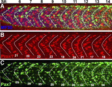
Fig. 5 Hh regulation of Pax7+ myogenic precursors occurs later than Hh induction of slow muscle fibers. Wild-type embryos were treated with cyclopamine from the 8-somite stage to the 30-somite (24 h) stage; they were then labeled for differentiated muscle fibers, slow muscle nuclei, and myogenic precursor nuclei. The numbers at the top of panel A indicate somite number. White lines frame the borders of somites: these were drawn solely with the MF20 labeling visible. (A) Merged image showing myosin (MF20, blue), slow muscle nuclei (Prox1, red), and myogenic precursor nuclei (Pax7, green). (B) A single color panel of A, showing Prox1-positive slow muscle nuclei in each somite, the number per somite is shown. All somites have an approximately wild-type number of Prox1-positive nuclei. Somites posterior to somite 18 had a loss of slow muscle fiber nuclei (data not shown, Hirsinger et al., 2004). (C) Single color panel of A, showing the Pax7-positive myogenic precursor nuclei in each somite. The number of Pax7+ myogenic precursors in somites 5 to 11 is similar to wild type, while posterior to somite 11, the number of Pax7+ myogenic precursors in each somite is increased to the level of smu-/-; compare to Fig. 2H. Anterior to the left, dorsal up; scale bar = 50 μm.
Reprinted from Developmental Biology, 300(2), Feng, X., Adiarte, E.G., and Devoto, S.H., Hedgehog acts directly on the zebrafish dermomyotome to promote myogenic differentiation, 736-746, Copyright (2006) with permission from Elsevier. Full text @ Dev. Biol.