
Fig. 2
- ID
- ZDB-IMAGE-070620-36
- Genes
- Publication
- Holtzman et al., 2007 - Endocardium is necessary for cardiomyocyte movement during heart tube assembly
- All Figures
- Figures for Holtzman et al., 2007
|
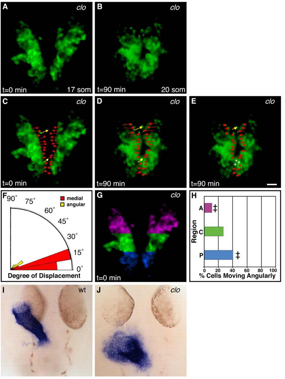
Fig. 2 No angular movement in the absence of endocardium. (A,B) Selected images from a time-lapse of cardiac fusion in a typical clo mutant zebrafish embryo expressing Tg(cmlc2:egfp) (see Movie 2 in the supplementary material), exhibiting cardiac morphology at the (A) 17-somite and (B) 20-somite stages. Dorsal views, anterior to the top. (C-E) Paths traveled during the (C,D) entire 90 minutes and (E) final 20 minutes of fusion (arrows and asterisks as described for Fig. 1). Tracks displayed represent a subset of the tracked cells in this time-lapse. In clo mutants, cardiomyocytes exhibit medial movement throughout the duration of cardiac fusion. Scale bar: in E, 20 μm for A-E. (F) Radial bar graph (see Fig. 1J,L) depicting degree of displacement for all tracked clo mutant cardiomyocytes. Few cells exhibit angular movement, resembling the pattern of cell behavior observed during the first phase of wild-type fusion (see Fig. 1J). (G,H) Location of cardiomyocytes moving angularly in clo mutants. In anterior (purple) and posterior (blue) regions (‡), the percentage of cells moving angularly is significantly reduced (t-test, P<0.01) compared with that observed during the second phase of wild-type fusion (see Fig. 1M). See Table 1 for additional data. (I,J) In situ hybridization depicts cmlc2 expression in wild-type (I) and clo mutant (J) embryos at 28 hpf. Dorsal views, anterior to the top; both images shown at the same magnification. Elongation of the heart tube is delayed and aberrant in clo mutant embryos.