
|
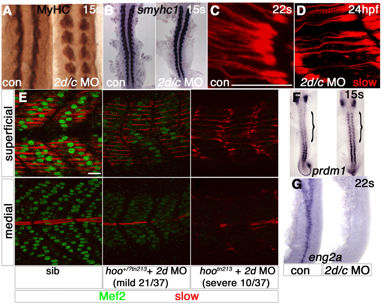
Fig. 3 Slow muscle myofibril defects correlate with the extent of Mef2 depletion. Zebrafish embryos in bright field (A,B,F,G), after immunodetection of all MyHC (A), slow MyHC (C-E) or Mef2 (E) or in situ mRNA hybridisation (B,F, dorsal flatmounts; G, lateral flatmount). (A,B) Slow muscle differentiation at 15 somites is unaffected by mef2d/c MO (A, somites 6-10). (C,D) Immature slow fibres in mef2d/c morphant at 24 hpf (D) are of comparable maturity to nascent fibres in a 20-hpf control embryo at the same rostrocaudal position (C). (E) Injection of mef2d MO into a hootn213 heterozygote cross yielded three phenotypes at the frequencies shown. Putative heterozygotes have diminished Mef2 in both slow (superficial slice) and fast (medial slice) fibres and thinner slow myofibril bundles. Putative mutants contained no Mef2 and had immature slow fibres. (F,G) mef2d/c morphants show persistence of prdm1 in adaxial cells (F, bracket) and loss of eng2a expression (G). Scale bars: 20 µm.