
|
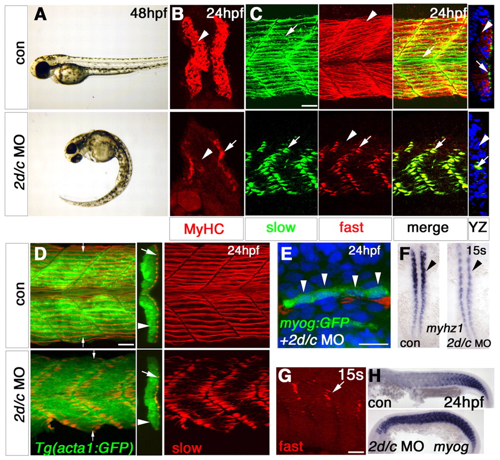
Fig. 2 Mef2 knockdown blocks fast muscle myofibril assembly. Zebrafish embryos in bright field (A,F,H), after immunodetection of all MyHC (B), slow MyHC (C-E) or fast MyHC (C,G) or in situ mRNA hybridisation (F, dorsal flatmount; H, lateral flatmount). (A,B) mef2d/c MO causes tail curvature (A) and ablates MyHC (B) in fast (arrowhead), but not slow (arrow) fibres. (C) mef2d/c morphants retain fast MyHC in slow fibres (arrows), but have essentially no fast MyHC in fast fibres (arrowheads), as shown in the yz optical transverse reconstruction. (D) Injection of mef2d/c MO into Tg(acta1:GFP) fish disrupts slow myofibrilogenesis (red) but not actin reporter expression (green). yz reconstruction at small arrows. Large arrows, slow fibres; arrowheads, fast fibres. (E) myogenin:GFP DNA co-injected with mef2d/c MO into wild-type embryos yields elongated GFP-filled fibres (green) with three to four nuclei (DAPI-stained, blue, arrowheads) at 24 hpf, despite disorganised slow MyHC (red). (F) Dorsal flatmounts showing decrease in myhz1 in fast precursors (arrowhead) but no change in adaxial slow precursors. (G) In wild-type embryos, fast MyHC is present in slow fibres at 15 somites (arrow) but is lost by 24 hpf (arrows, upper panels in C). (H) Persistence of myogenin mRNA in mef2d/c morphants. Scale bars: 20 µm.