
|
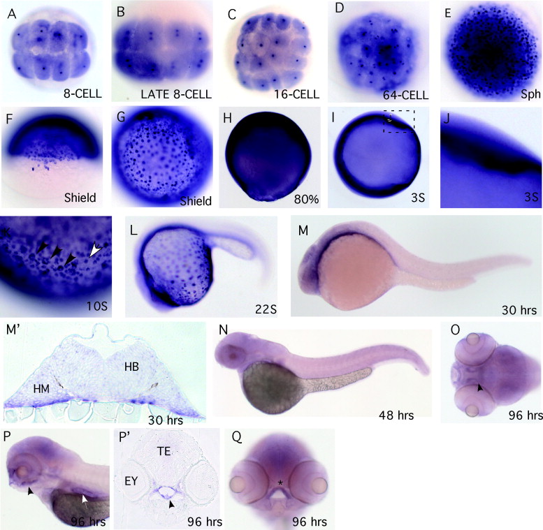
Fig. 2 Whole mount RNA in situ hybridization analysis of chch expression in zebrafish. The chch expression pattern was examined in zebrafish by RNA in situ hybridization. In situ hybridization probes to the 52-UTR and coding region or just the 32-UTR were used; both gave similar results and only one is shown. During cleavage stages, chch transcripts are detected in the cytoplasm of all cells. Concentrated stain is also detected in the nucleus of each cell (A–D). At sphere and shield stages, chch is widely expressed and some cells express higher levels of transcript including a subset of forerunner cells (E–G). At 80% chch is ubiquitously expressed throughout the embryo but the punctate stain is less apparent (H). From the three-somite stage through the 22-somite stage chch transcripts are weakly detected throughout the embryo with highest levels in the ventral-most regions of the embryo close to the yolk (I–L) and in a punctate pattern on the surface of the yolk (focus on the surface of the yolk) (K). In panel (K), the black arrowheads mark presumptive mucous cells and the white arrowhead is a presumptive keratinocyte. At 30 hpf chch transcripts are enriched in anterior neural tissue and ventral cells adjacent to the yolk (M) and (M2). The section in (M2) is at the level of the hindbrain. At 48 hpf chch expression is weak and indistinct (N). At 96 hpf chch transcripts are detected in the pharynx (black arrowhead), gut (white arrowhead) and ethmoid plate (asterisk) (O–Q, section in P2). (A–E and G) Animal pole views; (F and O) dorsal views; (H–N and P) lateral views; and (Q) frontal view. Abbreviations used are: hindbrain (HB), head mesoderm (HM), eye (Ey) and tectum (TE).
Reprinted from Gene expression patterns : GEP, 7(6), Londin, E.R., Mentzer, L., Gates, K.P., and Sirotkin, H.I., Expression and regulation of the zinc finger transcription factor Churchill during zebrafish development, 645-650, Copyright (2007) with permission from Elsevier. Full text @ Gene Expr. Patterns