
|
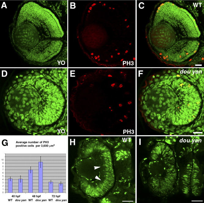
Fig. 6 The dou yanmi234 mutation does not disrupt cell proliferation in the retina.A-F: M-phase nuclei, visualized by anti-phospho-Histone 3 antibody (PH3, red), are observed in both wild-type (A-C) and mutant retinas (D-F) at 3 days postfertilization (dpf). All nuclei are stained with YO-PRO-1 iodide (YO, green). C is the merged image of A and B. F is the merged image of D and E. G: The average number of PH3-positive cells per 3,600 μm2 in mutant retinas is similar to wild-type retinas at 40 hours postfertilization (hpf), 48 hpf, and 72 hpf. The error bars denote the standard error of the mean. H: Bromodeoxyuridine (BrdU) labeling of the wild-type retina between 49 hpf and 51.5 hpf reveals proliferating cells in most regions of the retina. Arrowheads indicate BrdU-negative basal regions in the wild-type retina. I: BrdU labeling of dou yan mutants between 49 hpf and 51.5 hpf confirms that many retinal cells are actively synthesizing DNA. Scale bars = 20 μm.