
|
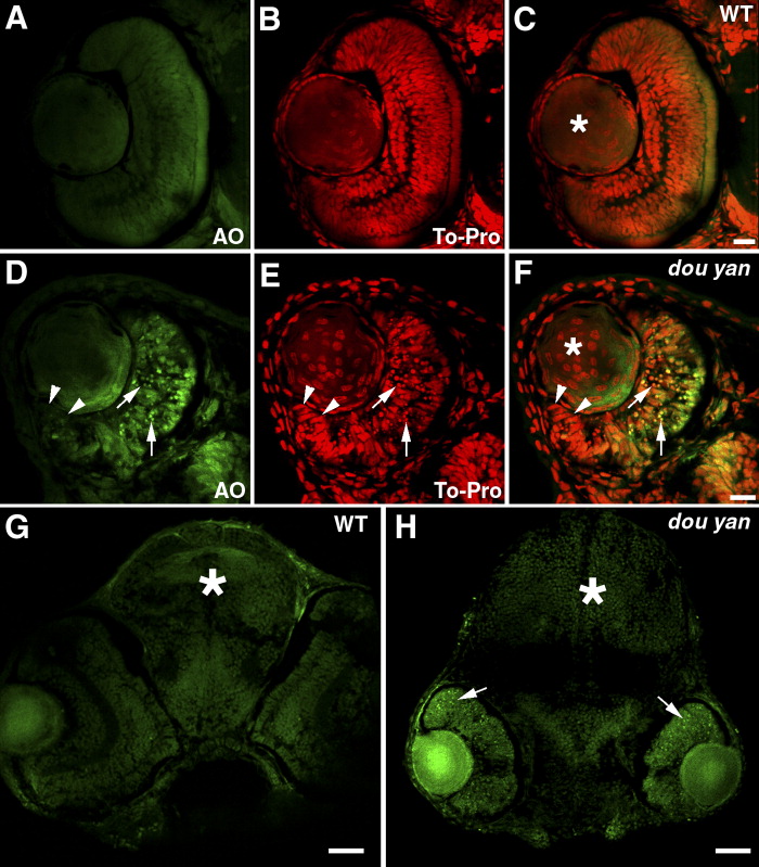
Fig. 4 The dou yanmi234 mutation causes extensive cell death in the retina but not in the brain or lens. A-F: Staining with acridine orange (AO, green), which preferentially stains the nuclei of dead or dying cells, indicates extensive cell death in the central regions of the mutant retinas (D-F, arrows) compared with the wild-type retinas (A-C). The cell nuclei are stained with TO-PRO-3 (red). Cell nuclei with an elongated shape are found at the marginal zone of the mutant retina (D-F, arrowheads), similar to the germinal zone of proliferating cells found at the retinal margin in wild-type retinas at this stage (A-C). In the lenses (asterisks), no increase in cell death was observed in dou yan. However, nonspecific acridine orange staining is sometimes observed in non-nuclear regions in the lens. G,H: Cell nuclei that are heavily stained with acridine orange are mainly found in the mutant retina (arrows) but not in the brain (asterisks). Scale bars = 20 μm in A-F, 50 μm in G,H.