
|
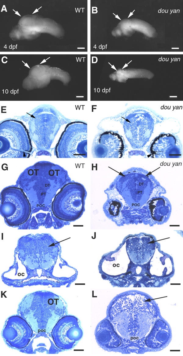
Fig. 3 The dou yanmi234 mutation preferentially affects the development of the optic tectum more than it does the other regions of the brain. A,B: Lateral views of the isolated wild-type (A) and mutant (B) brains at 4 days postfertilization (dpf) confirm that most regions of the mutant brain are only slightly smaller than those of the wild-type brain. However, the optic tectum is drastically reduced in the mutant embryos (arrows). C,D: The arrows indicate the optic tectum regions of brains isolated from wild-type (C) and surviving mutants (D) at 10 dpf. E,F: Cross-sections of the heads of wild-type (E) and mutant (F) embryos at the optic nerve region indicate similar forebrain size at 4 dpf, with the dorsal-ventral diameter of the mutant forebrains (arrows) being approximately 94% of that of the wild-type brains. The overall patterning of the gray (cell bodies and nuclei) and white (neuronal processes) matter is similar between the mutant and the wild-type embryos. Arrowheads indicate the optic nerves, which serve as a reference point for comparable section planes. G,H: The cross-sections of 4 dpf wild-type (G) and mutant (H) heads through the midbrain region reveal that the optic tectum (OT) is almost missing in the comparable region in the mutant embryos (arrows, H). However, the posterior commissure (POC), posterior tuberculum (PT), and the dorsal thalamus (DT) are comparable between mutant and wild-type embryos. I,J: The cellular patterning of the hindbrain mutant (J) is similar to that of the wild-type brain (I) at 4 dpf. Arrows indicate the hindbrains. The otic capsules (OC) were selected as a reference point for comparable section planes. K,L: At 3 dpf, cross-sections through the midbrain revealed the optic tectum in wild-type embryos (K). However, no optic tectum-like structure is evident in the mutant embryos (L) at the corresponding region (arrow). The posterior commissure (POC) is selected as a reference point for matching comparison. Scale bars = 100 μm in A,B,C-G, 50 μm in E,F,G-L.