
|
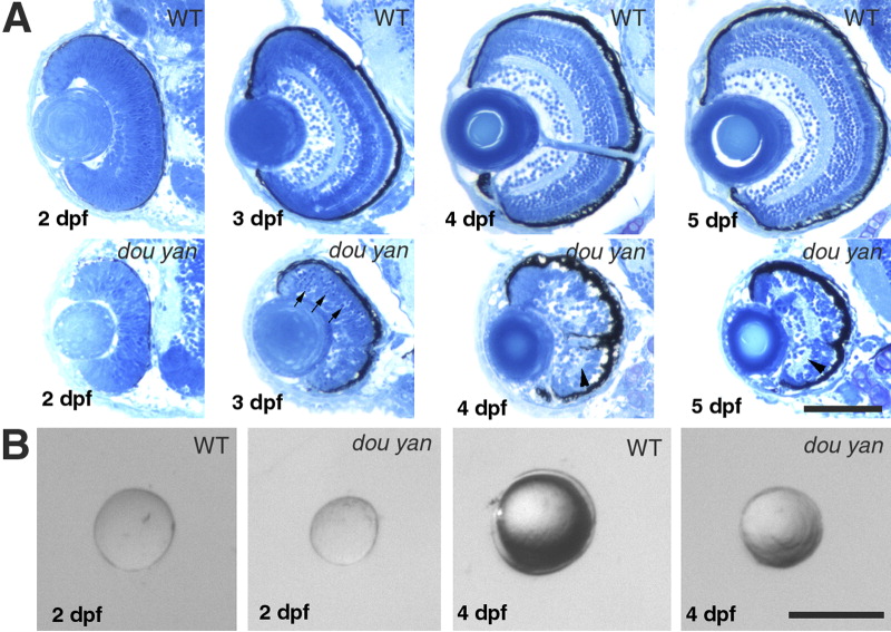
Fig. 2 The dou yanmi234 mutation disrupts cytoarchitecture of the retina and reduces the retinal size more severely than it does to the lens. A: Histological analyses sections of wild-type and mutant retinas at 2, 3, 4, and 5 days postfertilization (dpf) reveal that retinal lamination is severely disrupted in the mutant eyes. Arrows indicate the darkly stained pyknotic nuclei in the mutant retina. Arrowheads indicate the disrupted plexiform layers. B: The isolated lenses of mutant embryos are 6% and 30% smaller than their counterparts of wild-type embryos at 48 hours postfertilization (hpf) and 4 dpf, respectively. Scale bars = 100 μm.