
|
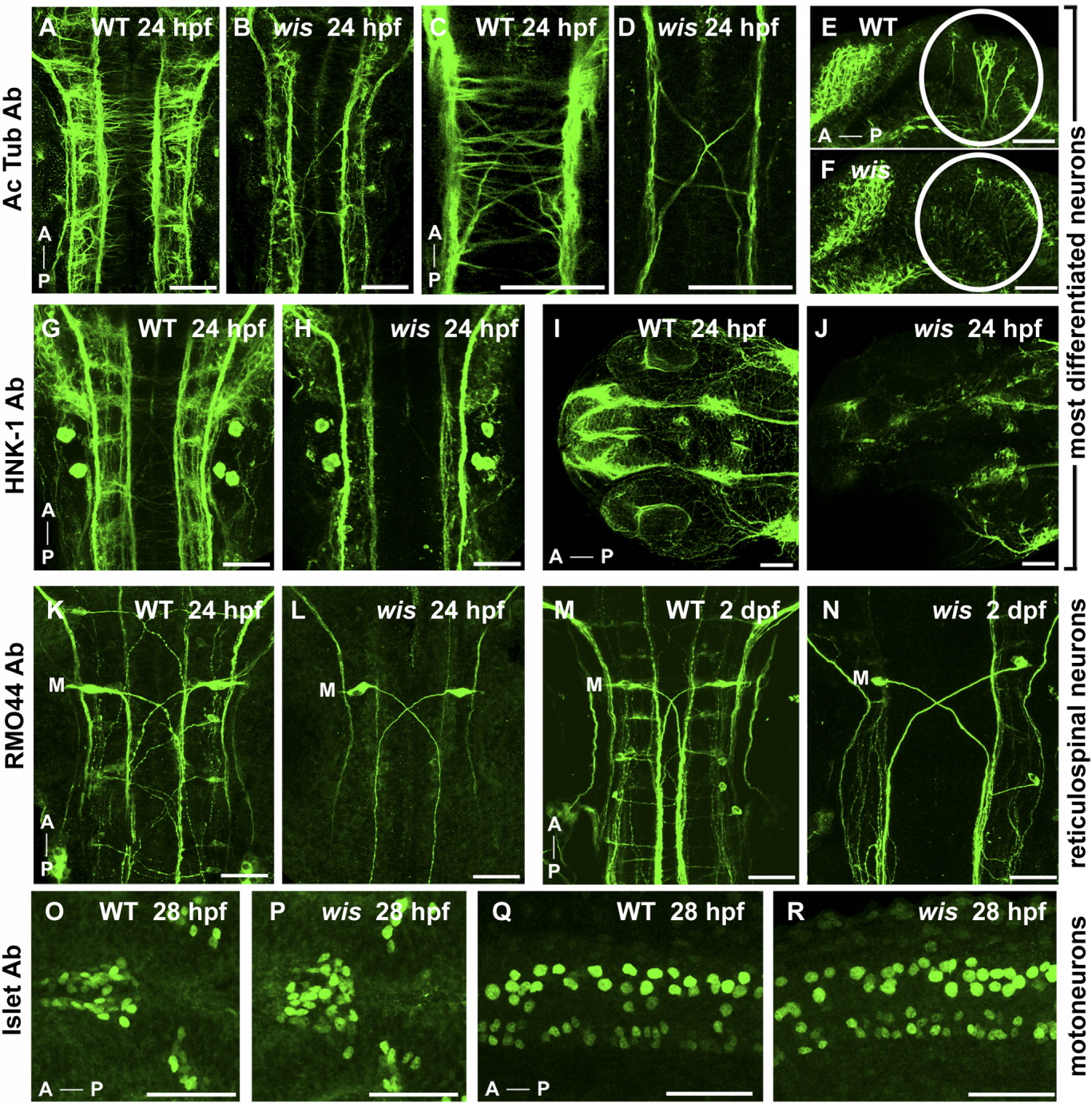
Fig. 7 Specific neurons are absent in whitesnake mutants. A-J: Acetylated tubulin antibody labeling (A-F) and HNK-1 antibody labeling (G-J), both of which mark most differentiated neurons and their axons, show reduced number of axons in wis mutant in the hindbrain (B,D,H), midbrain (F), and eyes/forebrain (J) compared with wild-type (A,C,E,G,I). K-N: The RM044 Ab, which labels reticulospinal neurons, shows an absence of all reticulospinal neurons in the wis hindbrain except Mauthner neurons (labeled with M) at both 24 hours postfertilization (hpf, L) and 2 days postfertilization (dpf, N), compared with wild-type (K,M). O-R: The 4D5 Islet antibody, labeling motoneurons, shows no loss of motoneurons in whitesnake in the dorsal diencephalon (P) or spinal cord (R) compared with wild-type (O,Q). A, anterior; P, posterior. A-D,G-P: Dorsal views. E-F,Q-R: Side views. Scale bar = 50 μm.