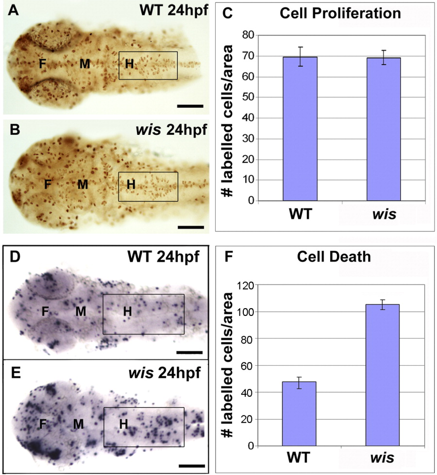
Fig. 5 Cell proliferation and cell death analysis in whitesnake mutants. A-C: Cell proliferation analysis, using PH3 antibody labeling. A,B: Fixed and labeled wild-type and wis brain at 24 hours postfertilization (hpf). C: Quantification comparing labeling in hindbrain shows no difference between wild-type and mutant, n = 8; P = 0.8046. D-F: Cell death analysis, using terminal deoxynucleotidyl transferase-mediated deoxyuridinetriphosphate nick end-labeling (TUNEL) staining. D,E: Fixed and labeled wild-type and wis brain at 24 hpf. F: Quantification comparing labeling in hindbrain shows approximately twice the amount of cell death in the mutant than in wild-type, n = 14; P < 0.0001. Error bars denote standard error. A-B,D-E: Dorsal views. Boxes mark regions used for quantitation. F, forebrain; M, midbrain; H, hindbrain. Scale bar = 100 μm.
Image
Figure Caption
Figure Data
Acknowledgments
This image is the copyrighted work of the attributed author or publisher, and
ZFIN has permission only to display this image to its users.
Additional permissions should be obtained from the applicable author or publisher of the image.
Full text @ Dev. Dyn.

