
|
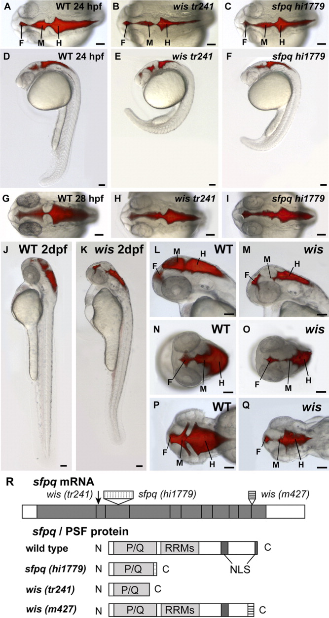
Fig. 1 Phenotype of whitesnake/sfpq mutants. A-Q: Brain ventricles were visualized by microinjecting a fluorescent dye, Rhodamine-dextran, into the hindbrain ventricle of living anesthetized embryos at 24 hours postfertilization (hpf, A-F), 28 hpf (G-I), and 2 dpf (J-Q). A-F: At 24 hpf, the wistr241 mutant has reduced brain ventricles and abnormal curvature of the tail (B,E), as compared with wild-type (A,D), and the sfpqhi1779 mutant phenotype is similar (C,F). G-I: At 28 hpf, both wistr241 and sfpqhi1779 show variable reduction in brain ventricle size and reduced pigmentation. J-Q: By 48 hpf, the wis brain ventricle reduction becomes more severe compared with wild-type, especially in the midbrain. A-C,G-I,N-Q: Dorsal views. D-F,J-M: Side views. F, forebrain; M, midbrain; H, hindbrain. Scale bar = 100 μm. R:sfpq gene/PSF protein and corresponding mutations. wistr241 has a C to T mutation at position 491 of coding sequence, which results in an early stop codon at amino acid 167. sfpqhi1779 has a 6-kb retroviral insertion (which has a stop codon early in the insertion sequence). wism427 mRNA has aberrant splicing resulting in 200 base pairs of intronic sequence inserted before the last exon. Both sfpqhi1779 and wistr241 are truncated near the end of P/Q-rich region, and wism427 lacks the last NLS. P/Q, proline/glutamine-rich region; RRMs, RNA recognition motifs; NLS, nuclear localization sequence.