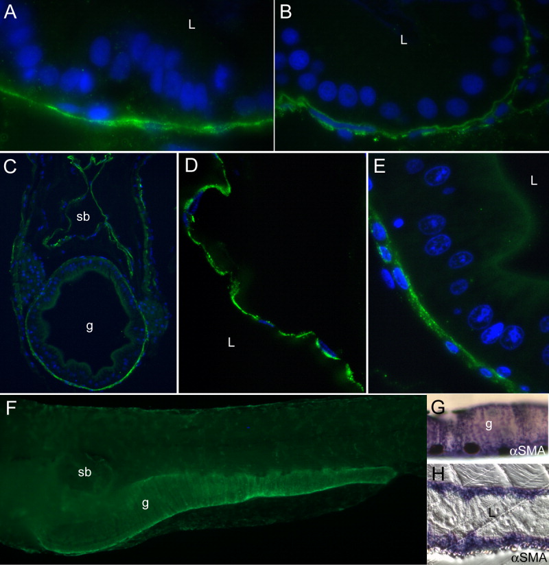
Fig. 5 Smooth muscle marker expression in the juvenile gut and swim bladder. A,B: Tropomyosin is expressed in a circular smooth muscle layer at 4 days postfertilization (dpf; A), and in longitudinal and circular layers at 6 dpf (B). C-F: By15 dpf, tropomyosin staining is strong in a layer surrounding both the gut and swim bladder (C). D,E: A higher power image (D) reveals that the swim bladder has a thin epithelial lining and single layer of smooth muscle cells surrounding it, while the gut (E) at similar magnification has developed a columnar epithelium, and a thicker smooth muscle wall. F: In a whole-mount larva, tropomyosin staining is seen around the swim bladder and gut. G: Whole-mount staining of a 20 dpf larva with α-smooth muscle actin (αSMA) reveals a similar pattern to tropomyosin. H: Longitudinal sections of a 22 dpf larva show extensive smooth muscle layers expressing αSMA. L, lumen; sb, swim bladder; g, gut.
Image
Figure Caption
Acknowledgments
This image is the copyrighted work of the attributed author or publisher, and
ZFIN has permission only to display this image to its users.
Additional permissions should be obtained from the applicable author or publisher of the image.
Full text @ Dev. Dyn.

