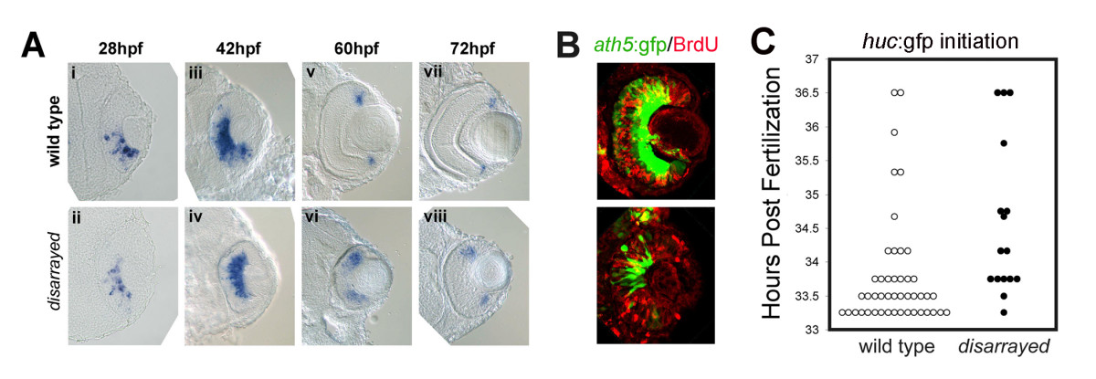
Fig. 4 Initiation of retinal neurogenesis in disarrayed eyes. (A) In situ analysis of ath5 expression during retinogenesis in wild-type (i, iii, v, vii) and disarrayed eyes (ii, iv, vi, viii). (B) BrdU pulse-labeling (red) in wild-type (upper) or disarrayed (lower) embryos carrying the ath5:gfp transgene (green). Embryos were injected with BrdU at 36 hpf and fixed at 42 hpf. Note that in both genotypes, ath5:GFP expression in central retinal cells is post-S-phase. (C) Developmental time (hours post fertilization) for the initiation of huc:GFP expression. The initiation of GFP expression was recorded for all embryos at 0.5 hr intervals in a clutch from an in-cross of disarrayed heterozygous parents (one circle represents one embryo; mutant (black circles) and wild-type (white circles)). Note that all embryos initiate huc:GFP expression within the same window of development. Results from one representative experiment (n = 16 mutant and n = 45 wild-type embryos from one clutch of embryos).
Image
Figure Caption
Figure Data
Acknowledgments
This image is the copyrighted work of the attributed author or publisher, and
ZFIN has permission only to display this image to its users.
Additional permissions should be obtained from the applicable author or publisher of the image.
Full text @ BMC Dev. Biol.

