
|
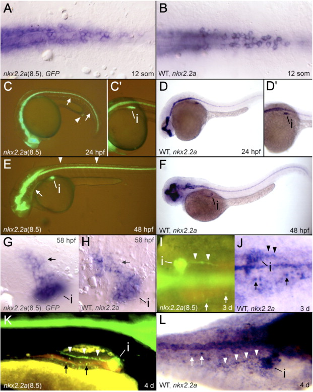
Fig. 1 Stable transgenic expression of GFP compared to nkx2.2a mRNA expression. (A, C, C2, E, G, I, K) GFP fluorescence or GFP mRNA expression in transgenic animals. (B, D, D2, F, H, J, L) nkx2.2a in situ hybridizations in WT zebrafish. (C–F, K) Lateral views. (L) Dorsolateral view. (I, J) Dorsal views. (A, B, G, H) Ventral views. (C2, D2) represent the same embryos shown in panels C and D respectively, focusing on pancreatic expression, dorso-lateral views. (C) Hypochord (arrows), proctodeum (arrowhead). (E) Branchial arches (arrow), dorsal spinal cord cells (arrowheads). (G, H) The arrow indicates GFP and nkx2.2a expression anterior and lateral to the pancreatic islet (i). (I–L) The pancreatic duct is indicated by arrowheads and enteroendocrine cells by arrows. i = pancreatic islet.
Reprinted from Developmental Biology, 304(2), Pauls, S., Zecchin, E., Tiso, N., Bortolussi, M., and Argenton, F., Function and regulation of zebrafish nkx2.2a during development of pancreatic islet and ducts, 875-890, Copyright (2007) with permission from Elsevier. Full text @ Dev. Biol.