
Image
|
Figure Caption
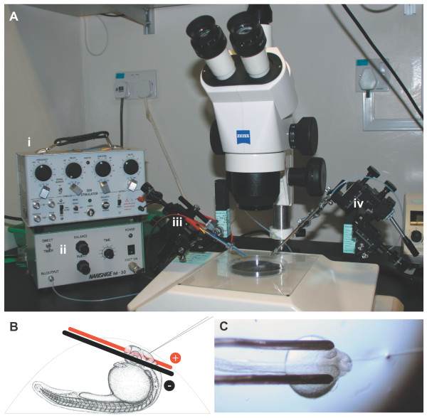
Fig. 1 Electroporation apparatus. (a) The electroporation equipment assembled on a dissecting microscope: the Grass SD9 stimulator (i) and air pressure injector (ii) are connected to two micromanipulators controlling the electrodes (iii) and microinjection needle (iv). (b) Side view schematic of a 1 dpf zebrafish mounted in an agarose drop with the electrodes and injection needle in position. Electrodes are not drawn to scale. (c) Top view of an embryo mounted for electroporation, with electrodes in position and microinjection needle inserted into the brain ventricle.
Acknowledgments
This image is the copyrighted work of the attributed author or publisher, and
ZFIN has permission only to display this image to its users.
Additional permissions should be obtained from the applicable author or publisher of the image.
Full text @ Neural Dev.