
|
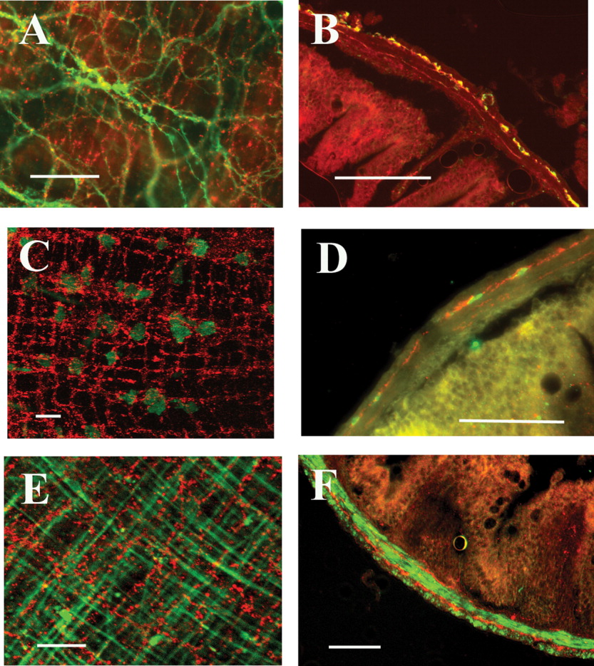
Fig. 3 Cells with Kit-like immunoreactivity form a dense network in the myenteric plexus region, and a second network close to the circular smooth muscle-submucosal border. High magnification of Kit-positive cells reveals individual stellate-shaped cells (white) in a full thickness stack (A). Circular and longitudinal muscle layers of the adult GI tract are clearly observed in H&E stained transverse sections (B). Kit-positive cells are found in the myenteric plexus region, and a thin layer of Kit-positive cells is located near the circular smooth muscle-submucosal border (C). A single layer of Kit-positive cells was observed in the tunica muscularis of transverse sections of zebrafish larvae (white arrow) (D). Scale bars = 20 μm.