
|
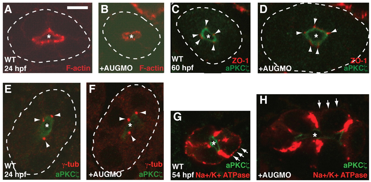
Fig. 7 Apical-basal distribution of Na+/K+-ATPase is disrupted in morphants. Cryosections through pronephric ducts of uninjected WT (A,C,E,G) and MOAUG-injected (B,D,F,H) embryos at the indicated developmental times. The lumen (apical) is indicated by an asterisk, and the basal sides of ducts are outlined by dotted lines in A-F. (A,B) Filamentous actin in the pronephric ducts (red). (C,D) Cell junctions are stained with ZO1 antibody (red, arrowheads), and the apical region of the cells is marked by aPKCζ (green). (E,F) Microtubule-organizing centers are stained with anti-γ-tubulin (red, arrowheads), and the apical region of the cells is marked by aPKCζ (green). (G,H) Localization of Na+/K+-ATPase (red); the apical region of the cells is marked by PKCζ (green). Arrows highlight the presence of staining in basal regions of WT control cells and its absence in morphants. Scale bar: 5 μm.