
|
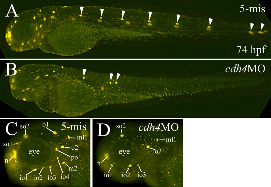
Fig. 7 DASPEI labeling of neuromasts in a 5-mis MO-injected embryo (A,C), and embryos injected with RcadMphA (cdh4MO, B,D). A and B are lateral views of whole live embryos (anterior to the left and dorsal up) with the same magnification. The neuromasts in the body and tail on the same side are indicated by arrowheads in these panels. C,D: Higher magnifications of the lateral view of the head region with anterior to the left and dorsal up. io1-4, infraorbital line neuromasts 1-4; n, nasal organ; m2, middle lateral line neuromast 2; ml1, middle line neuromast 1; o1 and o2, otic lateral line neuromasts 1 and 2; po, postorbital neuromast; so1 and so2, supraorbital line neuromasts 1 and 2.