
|
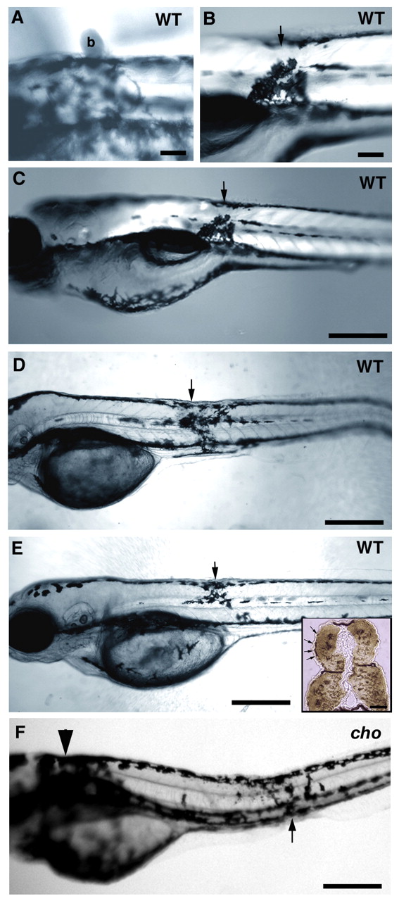
Fig. 9 Implantation of Sdf1-soaked beads into WT and cho mutant embryos results in the formation of an ectopic melanophore collar. (A) SDF1-soaked beads (b) were implanted in a dorsal position directly against the skin of a WT embryo at 24 hpf, held in place by a drop of low melting point agarose. Embryos were photographed at 48 hpf to ensure the bead had stayed in place and to record the location of the bead. Agarose holding the bead distorts light transmission, causing the halo evident in this image. (B) Same embryo as in A but at 72 hpf, after bead removal to document the phenotype. The position at which the bead was placed is marked with an arrow. Melanophores have accumulated ectopically, adjacent to the location of the bead implant. (C) Low magnification of same larva as in B. (D,E) Two independent WT embryos, treated similarly, also show ectopic melanophore accumulations adjacent to the location of the SDF1 bead (arrows). Ectopic melanophores form regardless of where in the embryo the bead is positioned. Insert in E is the same animal sectioned in an area containing ectopic melanophores, immunolabelled for MyHc (brown) and demonstrating that bead implantation did not affect muscle differentiation or maintenance. Note that beads are not surgically implanted, but merely placed on the skin; thus, no wound is formed. (F) Implantation of SDF1 beads in homozygous cho mutant embryos (note collar, arrowhead) results in similar accumulation of ectopic melanophores adjacent to site of bead (arrow). Scale bars: 100 μm in A,B; 350 μm in C-F; 25 μm in insert.