Image


                        Figure Caption


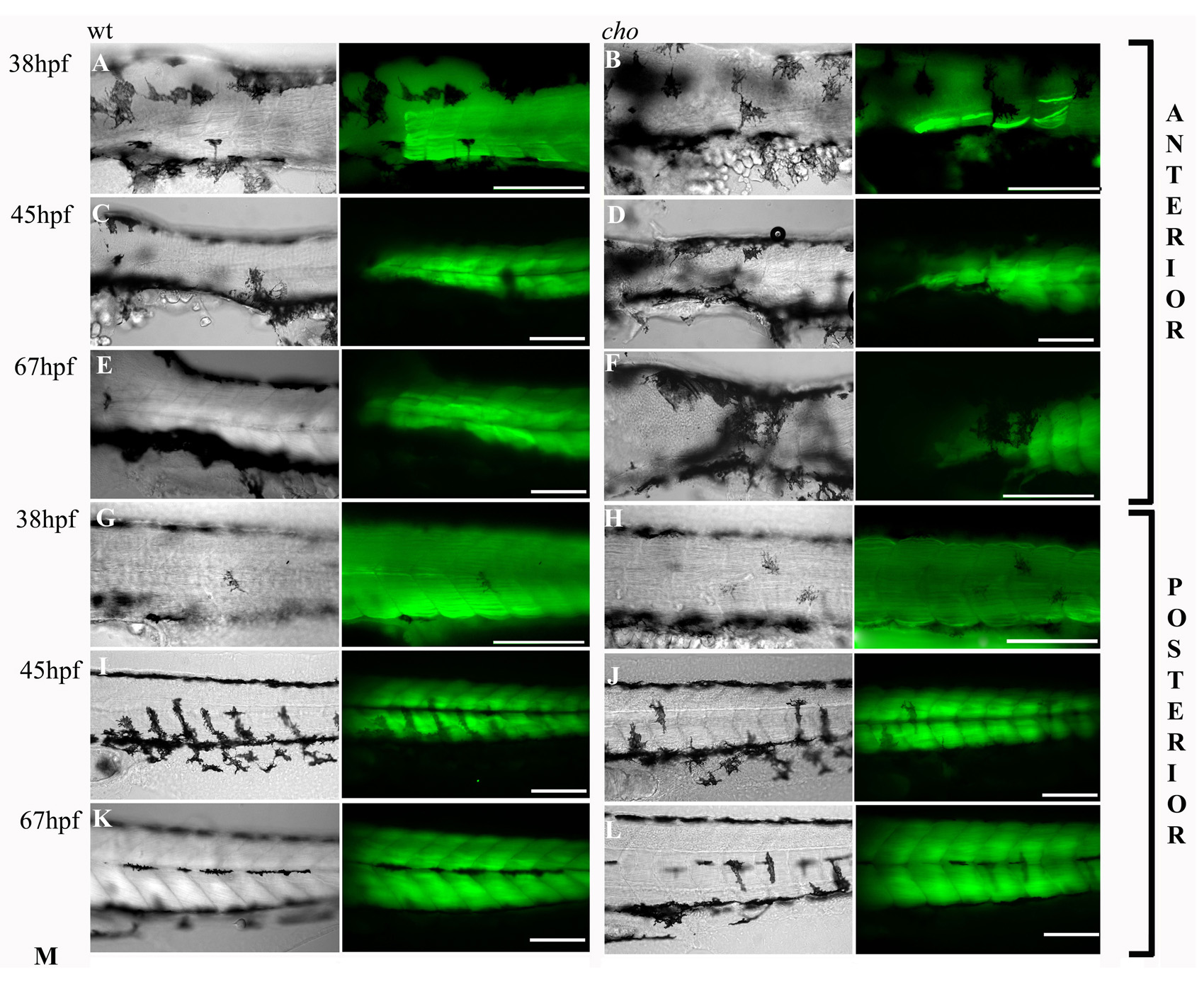
Fig. S1 Progressive nature of the muscle fibre defect in cho homozygotes. Slow muscle fibre arrangements and integrity as revealed by labelling with slow MyHc antibody (green). Left frame is a DIC image showing melanophore positions. The right frame combines this DIC image with a fluorescent image revealing F59 antibody labelling in one focal plane. Anterior trunk (A-F) and posterior trunk (G-L) are shown for WT (A,C,E,G,I,K) and cho mutant (B,D,F,H,J,L) embryos at 38 (A,B,G,H), 45 (C,D,I,J) and 67 hpf (E,F,K,L). Scale bars: 125 μm.
                
                    
                        Figure Data


                        Acknowledgments


This image is the copyrighted work of the attributed author or publisher, and
ZFIN has permission only to display this image to its users.
Additional permissions should be obtained from the applicable author or publisher of the image.
      Full text @ Development