
|
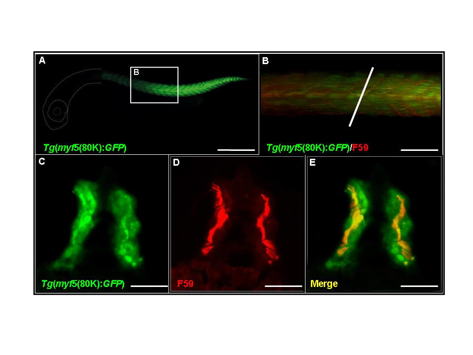
Fig. 3 Tg(myf5(80k):GFP) transgenic embryos express green fluorescent protein (GFP) in both slow and fast muscle fibers. (A,B) GFP expression in somites labeled with the F59 antibody. White dash lines indicate the location of head. (B) Higher magnification view of the boxed region shown in A. (C-E) Cross-section along the plane indicated by the white line in panel (B). GFP signals are observed in both fast (C, green signals) and slow muscle fibers (F, yellow signals). (A,B) Side views, rostral to the left, dorsal to the top; (C-D) dorsal to the top. 28 hpf. Scale bars: 400 μm in A; 200 μm in B; 100 μm in C-E.