
|
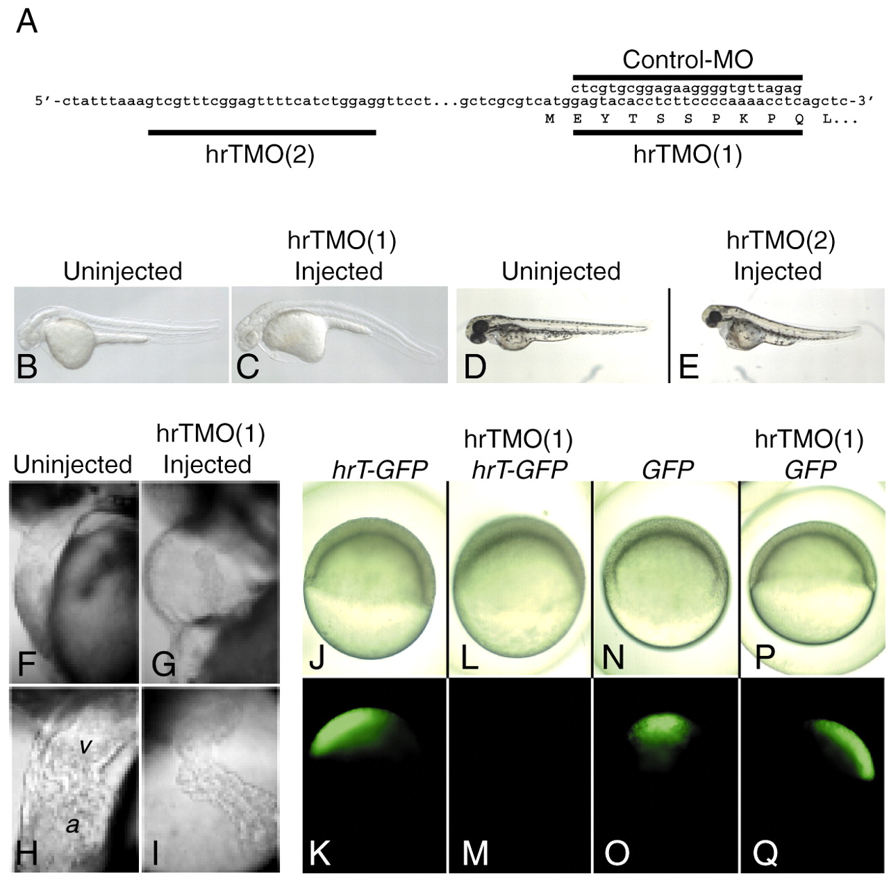
Fig. 1 hrT morphant phenotypes. (A) Binding positions of the morpholino antisense oligonucleotides, hrTMO(1) and hrTMO(2), to the hrT transcript. The sequence of the control-MO is also shown. (B-E) Lateral views with anterior towards the left. (B,C) The overall morphology of an uninjected (B) and hrTMO(1)-injected (C) embryos at 24 hpf. (D,E) The overall morphology of an uninjected (D) and hrTMO(2)-injected (E) embryos at 50 hpf. (F-I) Lateral view with anterior towards the top. (F,G) Heart morphology of uninjected embryo (F) and of hrTMO(1)-injected (G) embryo at 48 hpf. (H,I) Higher magnification of F,G, respectively (v, ventricle; a, atrium). Live (J,L,N,P) and green fluorescent (K,M,O,Q) pictures of embryos injected at the shield stage with: (J,K) 0.1 ng of hrT-GFP RNA; (L,M) 0.1 ng of hrT-GFP RNA plus 1.5 ng of hrTMO(1); (N,O) 0.1 ng of GFP RNA; and (P,Q) 0.1 ng of GFP RNA plus 1.5 ng of hrTMO(1).