
|
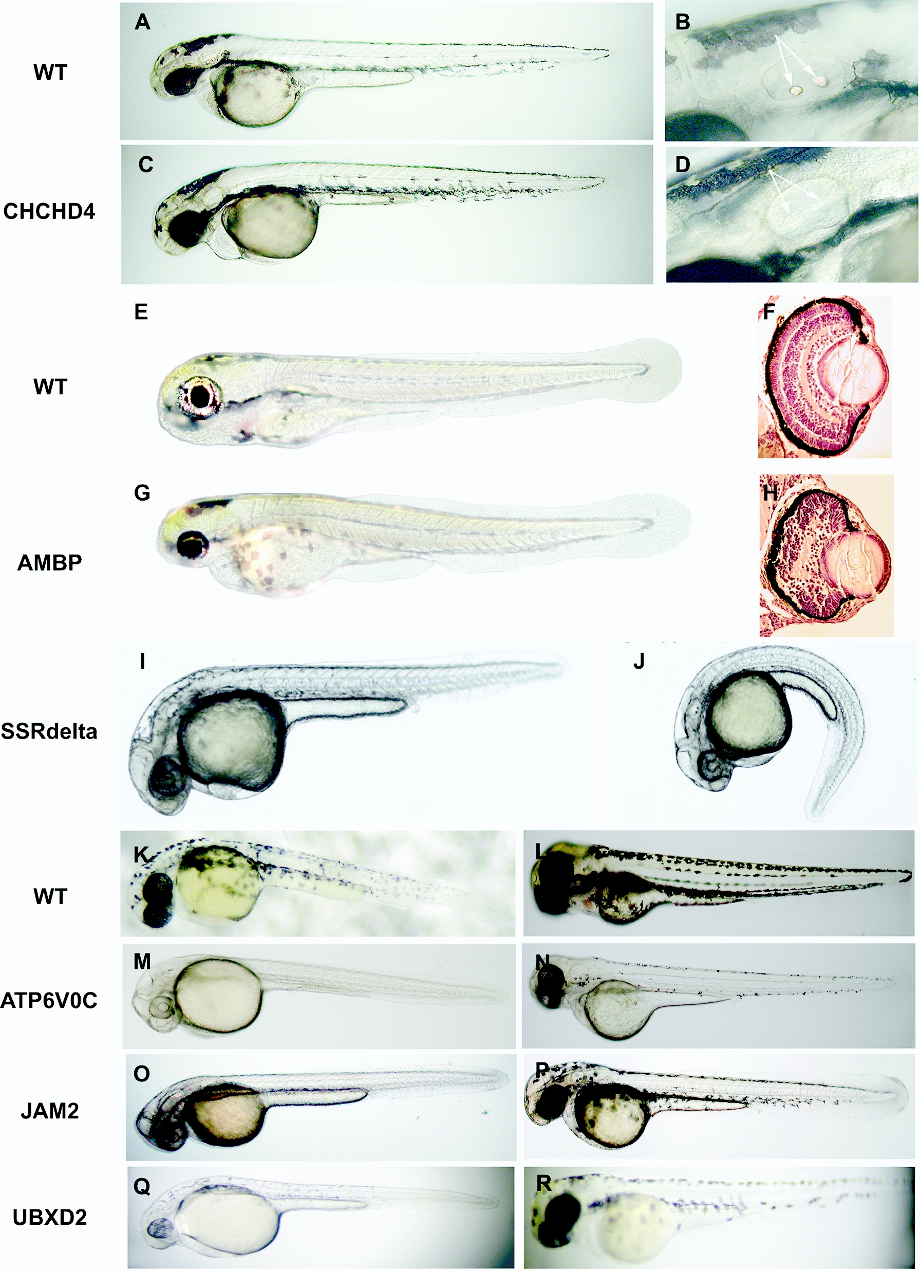
Fig. 5 Morphological defects observed following MO inactivation of select CTT genes. (A, B) Otolith morphology observed in 2 dpf untreated embryos. (C, D) Absence of otoliths, in otherwise normal 2 dpf embryo, following injection of MO targeting CHCHD4. (B, D) Enlarged view of otic capsules; arrows denote normally formed (B) or absent (D) otoliths, respectively. (E, F) Eye morphology in 3 dpf embryos. (G, H) Abnormally small eyes observed in 3 dpf embryo following injection of MO targeting AMBP. (F, H) Enlarged view of histological sections of eye in un-affected (F) and affected (H) embryos. Note differences in both the size and tissue organization of the affected eye. (I) Wild-type morphology of 1 dpf embryo. (J) Ventral curvature phenotype observed in 1 dpf embryos injected with MO targeting SSRdelta. (K, L) Normal pigmentation observed in untreated 1 and 2 dpf embryos. Reduction in pigment observed in 1 and 2 dpf embryos, respectively, following injection of MO targeting ATP6V0C (M, N)[23], or junction adhesion molecule 2 (JAM2) (O, P), or UBX domain containing 2 (UBXD2) (Q, R)).