
|
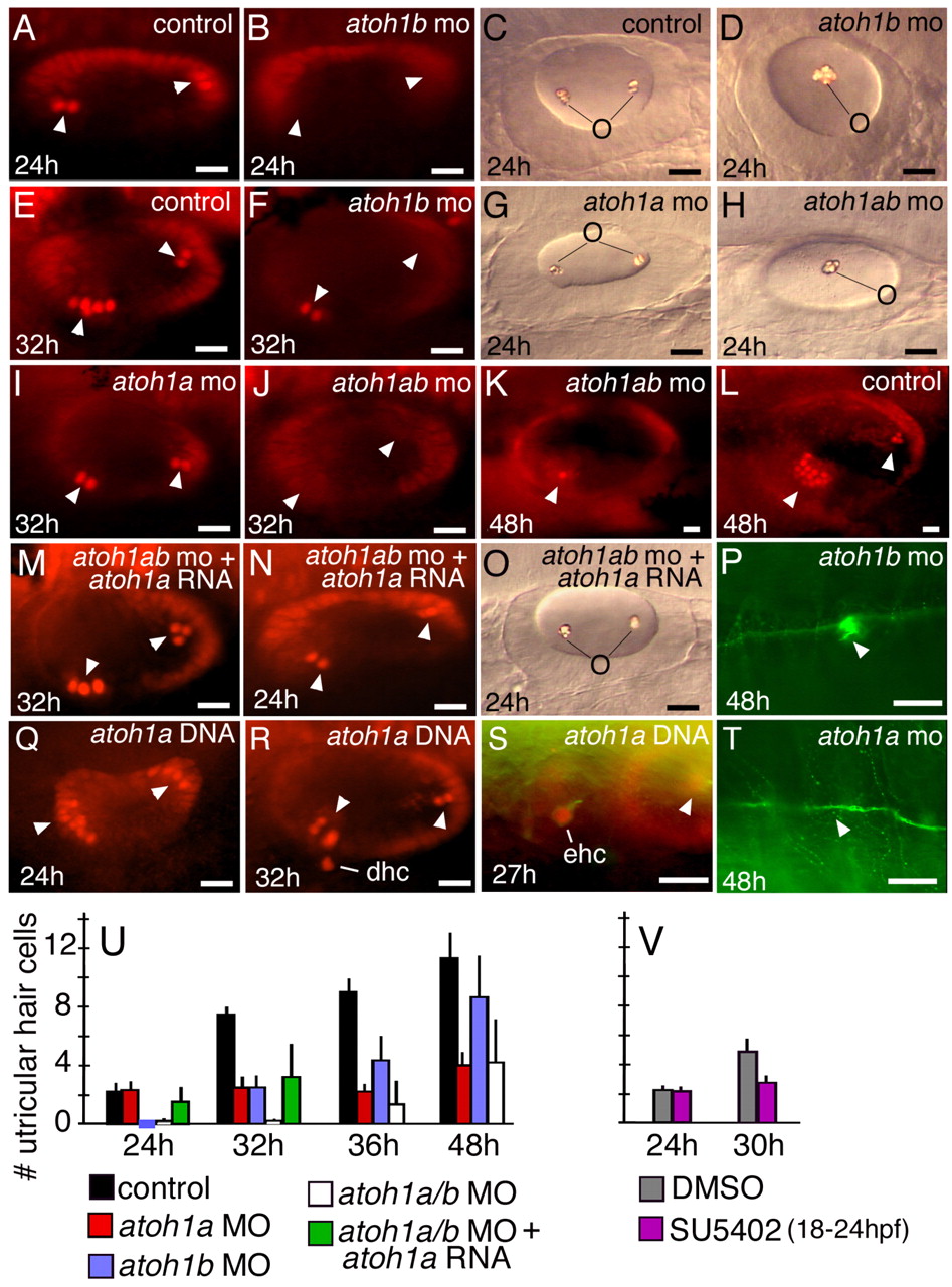
Fig. 2 Requirement for atoh1 in hair cells in the ear and lateral line. All panels show dorsolateral views with anterior to the left and dorsal up. (A,B,E,F,I-N,Q-S) Pax2 antibody staining of otic hair cells (arrowheads) at the indicated times in control embryos (A,E,L), atoh1a morphant (I), atoh1b morphants (B,F), atoh1a;atoh1b double morphants (J,K), atoh1a;atoh1b double morphant co-injected with atoh1a mRNA (M,N) and embryos injected with atoh1a plasmid (Q-S). atoh1a plasmid stimulates production of supernumerary hair cells at 24 hpf (Q), but these are not maintained at 32 hpf (R), and instead displaced hair cells appear ventrally within subjacent mesenchyme, leaving gaps in the hair cell layer. An ectopic hair cell is revealed anterior to the otic vesicle by co-staining with Pax2a (red) and acetylated-tubulin (green) (S). (C,D,G,H,O) Otoliths produced in control (C), atoh1a morphant (G), atoh1b morphant (D) atoh1a;atoh1b double morphant (H) and atoh1a;atoh1b double morphant co-injected with atoh1a RNA (O). (P,T) Acetylated-tubulin staining of the lateral line and neuromasts (arrowheads) in atoh1b morphant (P) and atoh1a morphant (T) at 48 hpf. (U,V) The mean (± standard deviation) of Pax2-postive hair cells present in the utricle at the indicated times and under the indicated conditions. Sample sizes ranged from 15-35 embryos per time point. Scale bar: 15 μm. dhc, displaced hair cells; ehc, ectopic hair cell; o, otolith.