
IMAGE
Fig. 2
Image
|
Figure Caption
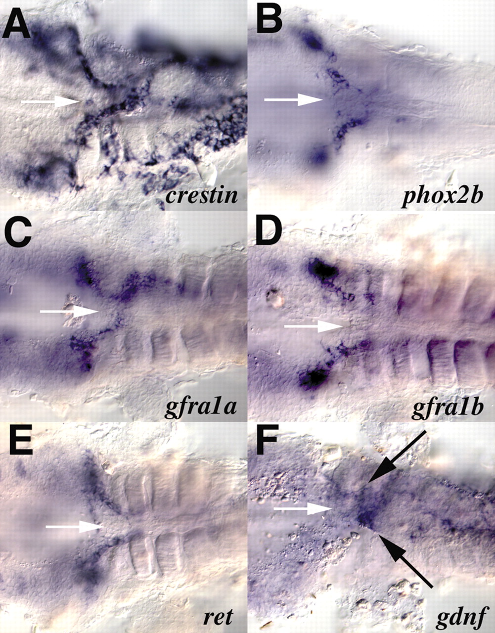
Fig. 2 Expression of gdnf receptor components in enteric precursors. Shown are ventral views of the vagal region of 36 hpf embryos after the yolk has been removed. Anterior is towards the left. In situ hybridization was performed for crestin (A), phox2b (B), gfra1a (C), gfra1b (D), ret (E) and gdnf (F). White arrows (A-E) indicate the anterior end of the gut tube. Black arrows in F indicate the mesendodermal expression of gdnf at the anterior end of the gut tube.
Figure Data
Acknowledgments
This image is the copyrighted work of the attributed author or publisher, and
ZFIN has permission only to display this image to its users.
Additional permissions should be obtained from the applicable author or publisher of the image.
Full text @ Development