
Fig. 3
|
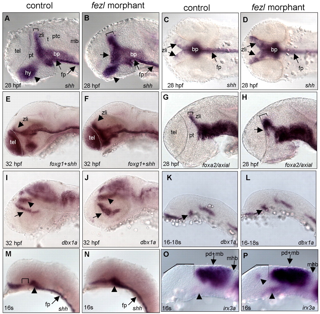
Fig. 3 The fezl morphants exhibit anterior expansion of the ZLI. (A-D) Marked anterior expansion of shh-expressing ZLI (B,D) was observed in fezl morphants. Arrows in A,B indicate the floor plate; arrowhead in A,B indicates hypothalamus; square brackets indicate the ZLI. (E,F) Double labeling of foxg1 (a telencephalic marker) and shh, showing the expression of shh in the prethalamic area in the fezl morphant. (G,H) Anterior expansion of foxa2 expression in the fezl morphant. Arrow in H indicates the expansion of foxa2-expressing cells; square brackets indicate the ZLI. (I,J) Expanded expression of dbx1a near the ZLI (arrow) and in the thalamus (arrowhead) in the fezl morphant. (K-P) dbx1a, shh and irx3a expression in the 16-somite-stage (16s) control and fezl morphants. Arrows in I-L indicate lhx5-expressing cells in the posterior tubercular region, and in M-N indicate the floor plate; arrowhead in I,J indicates lhx5-expressing cells in the thalamus, and in M-P indicates expanding shh expression in the presumptive ZLI; square bracket in M indicates the ZLI; dotted line in O,P indicates the anterior boundary of irx3a expression in the posterior diencephalon and midbrain region. All are lateral views of embryonic brains, except C,D, which are dorsal views. t, thalamus; hy, hypothalamus; mb, midbrain; ptc, pretectum; tel, telencephalon; fp, floor plate; bp, basal plate.