
Fig. 2
|
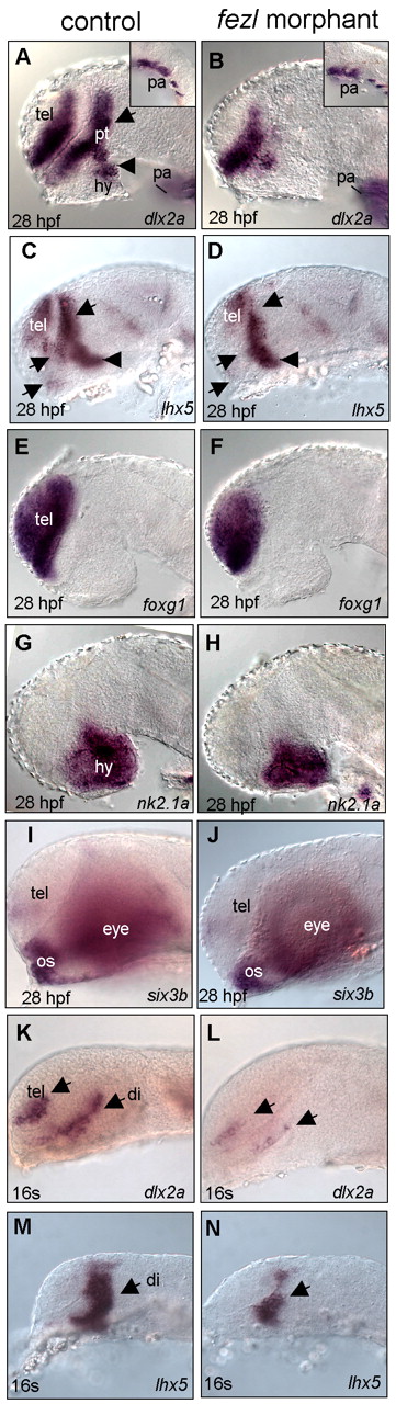
Fig. 2 Reduced fezl activity results in a deficit of the prethalamus. (A,B) Severe deficit of dlx2a in the prethalamus of a 28-hpf fezl morphant. Insets in A,B show unaffected dlx2a expression in the pharyngeal arch (pa) area. Arrow in A indicates dlx2a-expressing prethalamic cells; arrowhead in A indicates dlx2a-expressing hypothalamic cells. (C,D) Reduction of lhx5 in the prethalamic region of the fezl morphant. Arrows in C,D indicate lhx5-expressing prethalamic cells; arrowhead in C,D indicates lhx5-expressing posterior tubercular cells. (E-J) Expression of foxg1 in the telencephalon (E-F), nk2.1a in the hypothalamus (G-H) and six3b in the eye and optic stalk (I-J) are largely normal in fezl morphants. (K-N) Reduction of dlx2a and lhx5 expressions in 16-somite (16s) fezl morphants. Arrows in K,L indicate the telencephalon and prospective diencephalon; arrow in M,N indicates the prospective diencephalon. All are lateral views with specific stages and marker identities indicted in each panel. di, prospective diencephalon; hy, hypothalamus; pt, prethalamus; tel, telencephalon; os, optic stalk.