
IMAGE
Fig. 6
- ID
- ZDB-IMAGE-061227-24
- Publication
- Hu et al., 2006 - Egr1 gene knockdown affects embryonic ocular development in zebrafish
- All Figures
- Figures for Hu et al., 2006
Image
|
Figure Caption
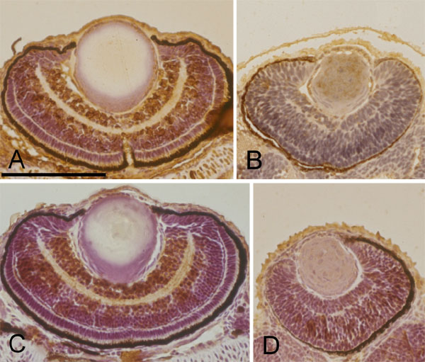
Fig. 6 Immunohistochemical staining of the wildtype and Egr1 morphants. Horizontal sections of zebrafish eyes at 72 h postfertilization with immunohistochemical stain for glutamate receptor 1 (A, B) and acetylated α-tubulin (C, D). Compared with the wildtype (A, C), retinal cells of the Grade-3 Egr1 morphant (B, D) arranged more compactly and disorderly. Significantly smaller areas of staining for both glutamate receptor 1 and acetylated α-tubulin appear in the morphant's retina. Scale bar represents 100 μm in panel A, and is applicable to all panels.
Acknowledgments
This image is the copyrighted work of the attributed author or publisher, and
ZFIN has permission only to display this image to its users.
Additional permissions should be obtained from the applicable author or publisher of the image.