
Fig. 5
- ID
- ZDB-IMAGE-061227-23
- Publication
- Hu et al., 2006 - Egr1 gene knockdown affects embryonic ocular development in zebrafish
- All Figures
- Figures for Hu et al., 2006
|
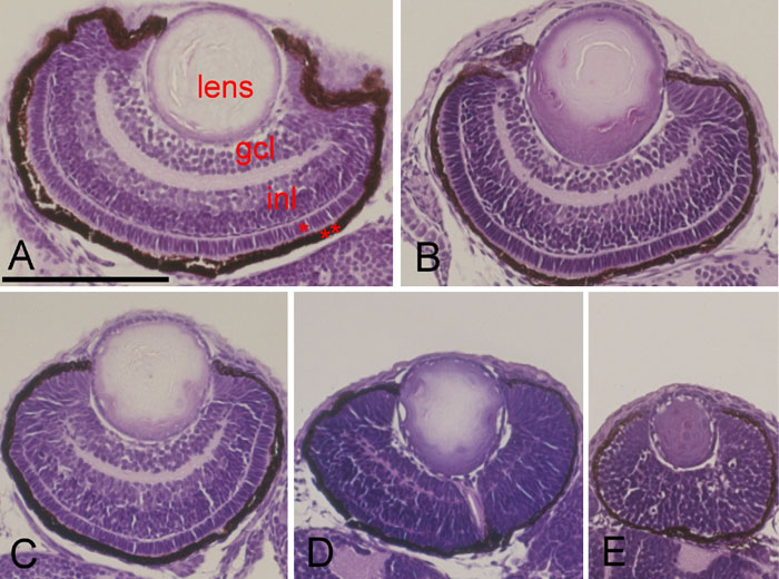
Fig. 5 Histological examination of the wildtype and Egr1 morphants. Hematoxylin and eosin stained transverse sections of zebrafish eyes at 72 h postfertilization having A: wildtype, B: Grade 1, C: Grade 2, D: Grade 3, or E: Grade 4. Higher-grade morphants have smaller eyes and smaller lenses. Both the inner plexiform layer between the ganglion cell layer (gcl) and inner nuclear layer (inl) as well as the outer plexiform layer between the inl and outer nuclear layer (*) are thin. The outermost retinal pigment epithelium (**) layer is also thin and irregular. Scale bar represents 100 μm in photo A, and is applicable to all photos.