
|
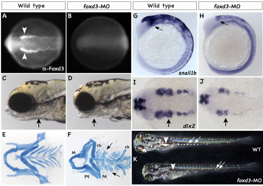
Fig. 8 Knockdown of the Foxd3 protein phenocopies mosm188 defects. A,B: Dorsal views of 4-somite stage embryos stained by immunohistochemistry using a zebrafish specific anti-Foxd3 antibody. In wild-type embryos (A), Foxd3 protein is localized to two parallel stripes of neural crest precursors at the edge of the neural tube (arrowheads). This pattern is lost in the foxd3 morphants (wild-type embryos injected with a cocktail of two foxd3 morpholinos) corroborating the effective and complete block of foxd3 translation (B). C,D: Lateral views of a live wild type (C) and a foxd3 morphant (D) at 5 dpf. The morphant phenotype closely resembles mosm188 mutant embryos with a gaping jaw and loss of posterior arches (arrows). E,F: Flat mounted Alcian blue stained cartilage preparations of a wild type (E) and a foxd3 morphant (F) show normal Meckel's (M) and palatoquadrate cartilages (pq). The second arch hyosymplectic (hs) in most cases is missing the hyomandibular portion. The ceratohyals (ch) are present, but the posterior pharyngeal arch cartilages are reduced to a few clusters of chondrocytes (arrows). The ceratobranchial (cb) cartilages are malformed. G,H: Expression of snail1b in neural crest precursors (arrows) is greatly reduced in the morphants (H). I,J: This result is concordant with reduced expression of migratory neural crest labeled with dlx2 (arrow points at second neural crest stream). K: Images of live wild-type and morphant embryos under incident light show overall reduced number of fluorescing iridophores in the lateral patch (arrowhead) and in the trunk (arrows) of morphants.