
|
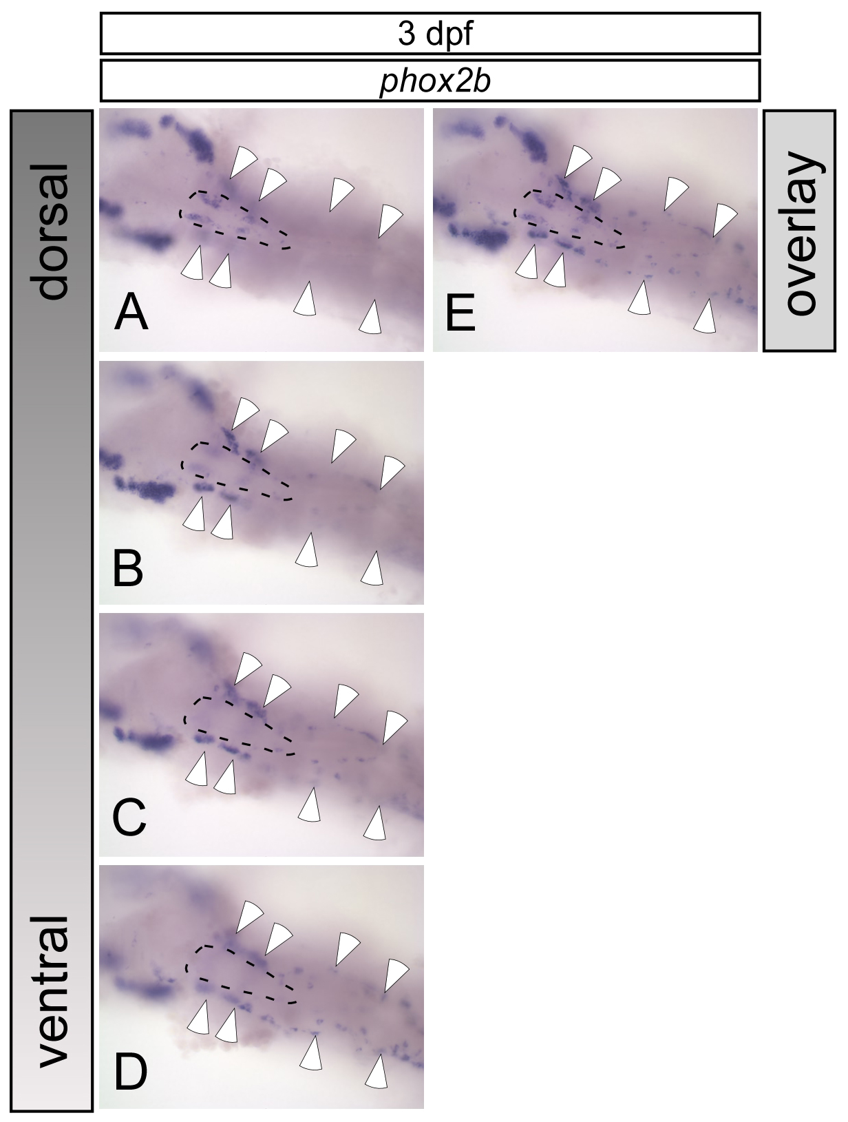
Fig. S2 Localisation of cervical sympathetic and enteric neurons in 3 dpf zebrafish embryos. (A-D) Whole-mount in situ hybridisation for phox2b as marker for autonomic cells was carried out. After dissection of dorsal hindbrain/spinal cord and yolk sac, a stack of pictures was made focussing from dorsal (A) to ventral (D) through the embryo. This illustrates the dorsal location of the cervical sympathetic ganglion (dashed circle), restricted to a relatively short anteroposterior area (A). Enteric neurons (white arrowheads) that appear as diffuse staining in A show up in more ventral focal planes (B-D), and extend to more rostral and caudal regions down to the anus (not shown). (E) An optical overlay of images in A to D to illustrate the different lateral positions of enteric and cervical sympathetic ganglion cells.