
|
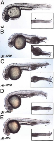
Fig. 4 Loss of ventral tail fin in din mutants. (A) Wild type embryos at 1dpf have a single ventral tail fin (arrows in ventral view inset). (B) A strong dintt250 mutant embryo at 1 dpf displays multiple ventral tail fins (white arrowheads). (C) Occasional dintt250 embryos display gaps in the multiplicated ventral fin (gap, black arrowheads; ventral fins, white arrowheads). dinm52 embryos also display alterations in the fin structure. (D) Most dinm52 embryos display a full ventral tail fin with minor duplications (white arrowheads) (E) Some embryos display reduced fins (black arrowhead) or gaps, in addition to duplications (white arrowheads). All panels lateral views, ventral view in inset.
Reprinted from Developmental Biology, 245(1), Wagner, D.S., and Mullins, M.C., Modulation of BMP activity in dorsal-ventral pattern formation by the Chordin and Ogon antagonists, 109-123, Copyright (2002) with permission from Elsevier. Full text @ Dev. Biol.