
Image
|
Figure Caption
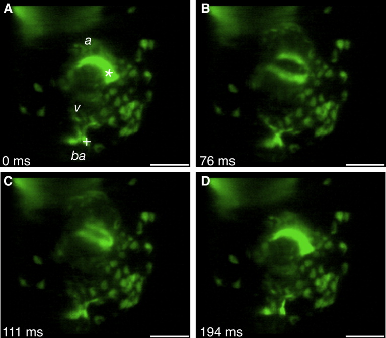
Fig. 4 Cardiac cushion dynamics during heart contraction. A-D: Single images produced from a four-dimensional reconstruction of the actively contracting 72 hours postfertilization (hpf) Tg(tie2:GFP)-expressing heart. The valve cushions between the atrium (a) and the ventricle (v) and the ventricle and the bulbus arteriosis (ba) are marked with an asterisk and plus sign, respectively. GFP, green fluorescent protein. Scale bar = 50 μm.
Acknowledgments
This image is the copyrighted work of the attributed author or publisher, and
ZFIN has permission only to display this image to its users.
Additional permissions should be obtained from the applicable author or publisher of the image.
Full text @ Dev. Dyn.