
|
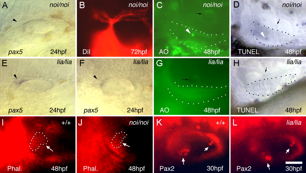
Fig. 7 Otic development in noi (pax2a) and lia (fgf3) mutants. A,E,F: Otic expression of pax5 in noi/noi (A) and lia/lia (E,F) mutants at 24 hours postfertilization (hpf). Arrowheads mark the pax5 expression domain. B: Statoacoustic ganglion (SAG) projections in a noi/noi mutant labeled by injecting DiI (1,1′, di-octadecyl-3,3,3′,3′,-tetramethylindo-carbocyanine perchlorate) into the utricular macula at 72 hpf. C,G: Acridine orange (AO) staining in noi/noi (C) and lia/lia (G) mutants at 48 hpf. D,H: terminal deoxynucleotidyl transferase-mediated deoxyuridinetriphosphate nick end-labeling (TUNEL) staining in noi/noi (D) and lia/lia (H) mutants at 48 hpf. White arrowheads indicate apoptotic cells in C and D, and black arrows mark otoliths. Utricular maculae are outlined in C, D, G, and H. I,J: Rhodamine-phalloidin labeling of the utricular macula (outlined) in wild-type (I) and noi/noi (J) embryos at 48 hpf. K,L: Anti-Pax2 staining of the otic vesicle in wild-type (K) and lia/lia (L) embryos at 30 hpf. I-L: White arrows indicate hair cell patches. A-L: Images show dorsolateral (A,B,E,F,I-L) and lateral (C,D,G,H) views, with anterior to the left. Scale bar = 30 μm in A,E,F,K,L, 50 μm in B, 25 μm in C,D,G,H,I,J.