
|
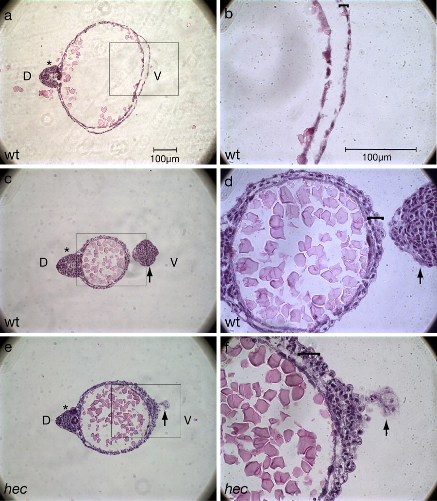
Fig. 5 Histological sections confirm the presence of additional cell layers in the yolk extension region. a-f: Hematoxylin/eosin labeling of a wild-type embryo (a-d) and a hec mutant embryo with a rudimentary axis (e,f). b,d, and f are magnifications of the boxed areas in a, c, and e, respectively. a,b: Cross-section within the region of the yolk cell proper of a wild-type embryo at the 18-somite stage (18 hpf). The wild-type yolk cell is bounded by a highly flattened epithelium (bracket in b). c,d: Cross-section within the yolk extension region of the same wild-type embryo as in a,b. d: The wild-type yolk extension is surrounded by a surface consisting of multiple layers of less flattened and often protruding cells (bracket). e,f: Cross-section within the region of embryonic constriction of a partial axis hec mutant embryo at 18 hours postfertilization (hpf). f: Like the yolk extension in wild-type embryos, the ventral yolk surface of partially ventroposteriorized hec mutant embryo is bounded by a surface consisting of several cuboidal cells (bracket). This surface, however, typically consists of more layers than in the yolk extension region of wild-type embryos. The tail (indicated by a black arrow) curves downward at this stage so that it is also visible in sections c-f. In a,c, the embryonic axis (or rudimentary axis in e) is indicated with an asterisk, and "D" and "V" in a,c denote the dorsal and ventral regions, respectively (also indicated in the axis-deficient hec mutant embryo in e as deduced by the presence of the rudimentary dorsal axis).