
|
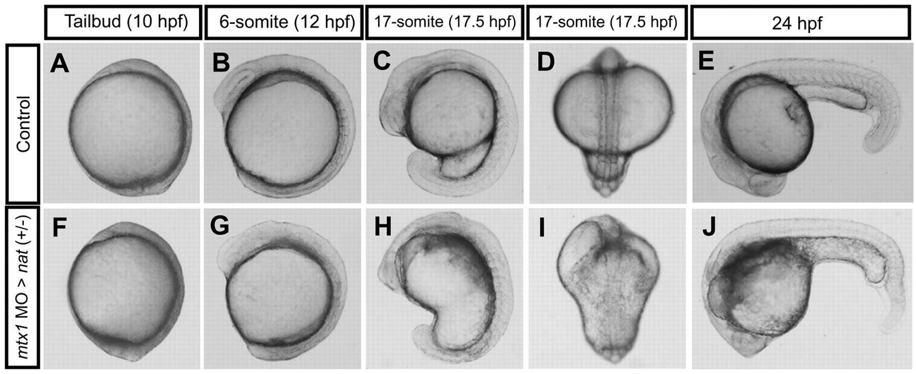
Fig. 6 Morphogenesis of mtx1 MO-injected heterozygous natter embryos. (A-E) Wild-type embryo; same embryo is shown at all stages. (F-J) heterozygous natter embryo injected with mtx1 MO(A) into the YSL at the 1000-cell stage; same embryo is shown at all stages. Lateral views, animal pole up and dorsal to the right (A-C,F-H); dorsal views, animal pole up (D,I), lateral views, anterior to the left and dorsal up (E,J). mtx1 MO-injected heterozygous natter embryo showed no apparent defect at the tailbud stage (A,F). A phenotype first appeared at the 6-somite stage at the hindbrain level as the YSL got darker (G). At the 17-somite stage, the head ventricle was almost collapsed onto the YSL (I). At 24 hpf, somite boundaries are clearly visible in wild-type embryos (E), whereas they were harder to visualize in mtx1 MO-injected heterozygous natter embryos (J), especially in the anterior part.