
|
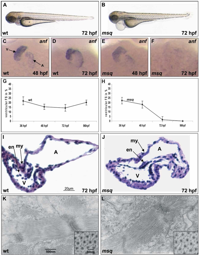
Fig. 1 Effects of the msq m347 mutation on heart function, morphology, and expression of anf. (A,B) Lateral view of wild-type (wt) (A) and msq mutant embryos (B) at 72 hpf. msq mutants develop pericardial edema due to loss of ventricular contractility, whereas the development of other organ systems proceeds normally. (C–F) RNA levels of the stretch-responsive gene zanf are severely reduced in msq mutant hearts. In contrast to wild-type hearts, where robust expression of zanf throughout the heart can be observed at 48 hpf (C) and 72 hpf (D), zanf expression in msq mutant hearts is only slightly reduced at 48 hpf (E), but completely absent by 72 hpf (F). Atrium (A) and ventricle (V) are indicated. (G,H) Contractility of msq mutant zebrafish embryo ventricles severely decreases over time; by 96 hpf, the ventricular chamber becomes completely silent. Measurements of fractional shortening (FS)—an indicator of systolic contractile function normalized to the diameters of the heart—are displayed for the ventricular chamber of wild-type zebrafish embryos (G) and msq mutants (H) at different time points. (I,J) The msq mutation does not affect overall heart morphology. Hematoxylin/eosin-stained histological sections of wild-type (I) and msq mutant (J) heart at 72 hpf. Endocardial (en) and myocardial (my) layers of ventricle and atrium of msq mutant heart are unaltered. (K,L) Transmission electron microscopy of wild-type (K) and msq mutant (L) zebrafish embryonic hearts at 72 hpf. Ventricular cardiomyocytes of msq mutants show normal cell architecture and organization of myofibrils. Insets display transverse sections of myofilaments.