
Image
|
Figure Caption
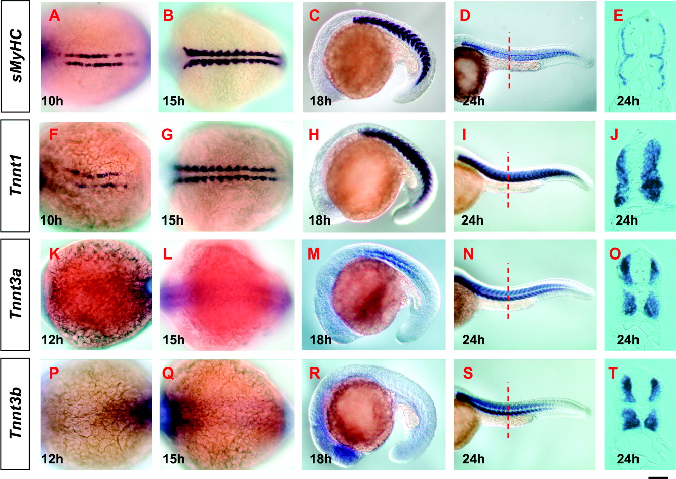
Fig. 7 Whole-mount in situ hybridization of Tnnt1, Tnnt3a, Tnnt3b, and sMyHC during zebrafish early somitogenesis. Embryos were hybridized with sMyHC (A-E), Tnnt1 (F-J), Tnnt3a (K-O), and Tnnt3b (P-T) riboprobes, respectively. A,B,F,G,K,L,P,Q: Dorsal views. C,D,H,I,M,N,R,S: Lateral views. E,J,Q,T: Transverse sections at trunk level. The anterior is to the left in all whole-mount stained embryos. Dotted lines indicate the section level. Embryonic stages are indicated in each panel. h, hour. Scale bar = 200 μm in D,I,N,S, 140 μm in C,H,M,R, 100 μm in A,B,F,G,K,L,P,Q, 25 μm in E,J,O,T.
Figure Data
Acknowledgments
This image is the copyrighted work of the attributed author or publisher, and
ZFIN has permission only to display this image to its users.
Additional permissions should be obtained from the applicable author or publisher of the image.
Full text @ Dev. Dyn.