
|
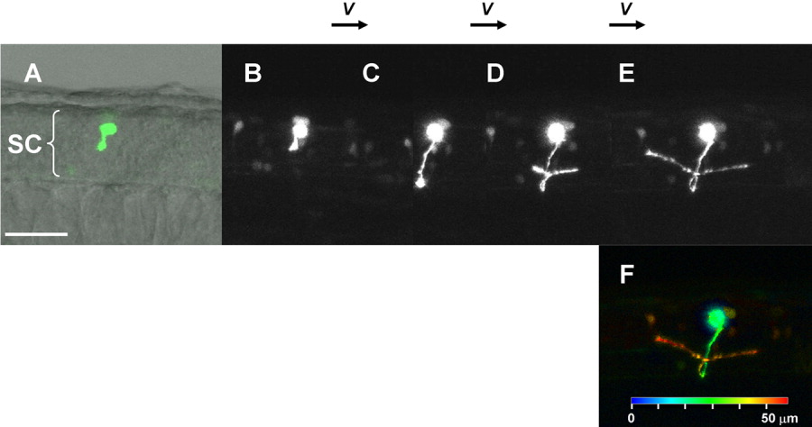
Fig. 4 Time-lapse imaging of neural development using Dronpa. A: A single neuron expressing Dronpa starting to extend axon at 27 hours postfertilization (hpf). Anterior is to the left. B-E: Time-lapse series of axonal navigation of the T-shaped commissural spinal neuron imaged at 27 hpf (B), 31 hpf (C), 39 hpf (D), and 42 hpf (E). The axon extended ventrally (B), crossed the midline (C), reached the contralateral side, and bifurcated both anteriorly and posteriorly (D). The neuron was irradiated with the 405-nm laser, immediately after every three time points for observation. This strategy resulted in recovery of the weakening fluorescence caused by observation using the laser with the same wavelength for the elimination. F: Depth pseudo-color code representation of the neuron at 42 hpf, showing the three-dimensional structure of the neuron. The scale for the depth is shown at the bottom. See also Supplementary Movie S4. Scale bar = 50 μm.