
|
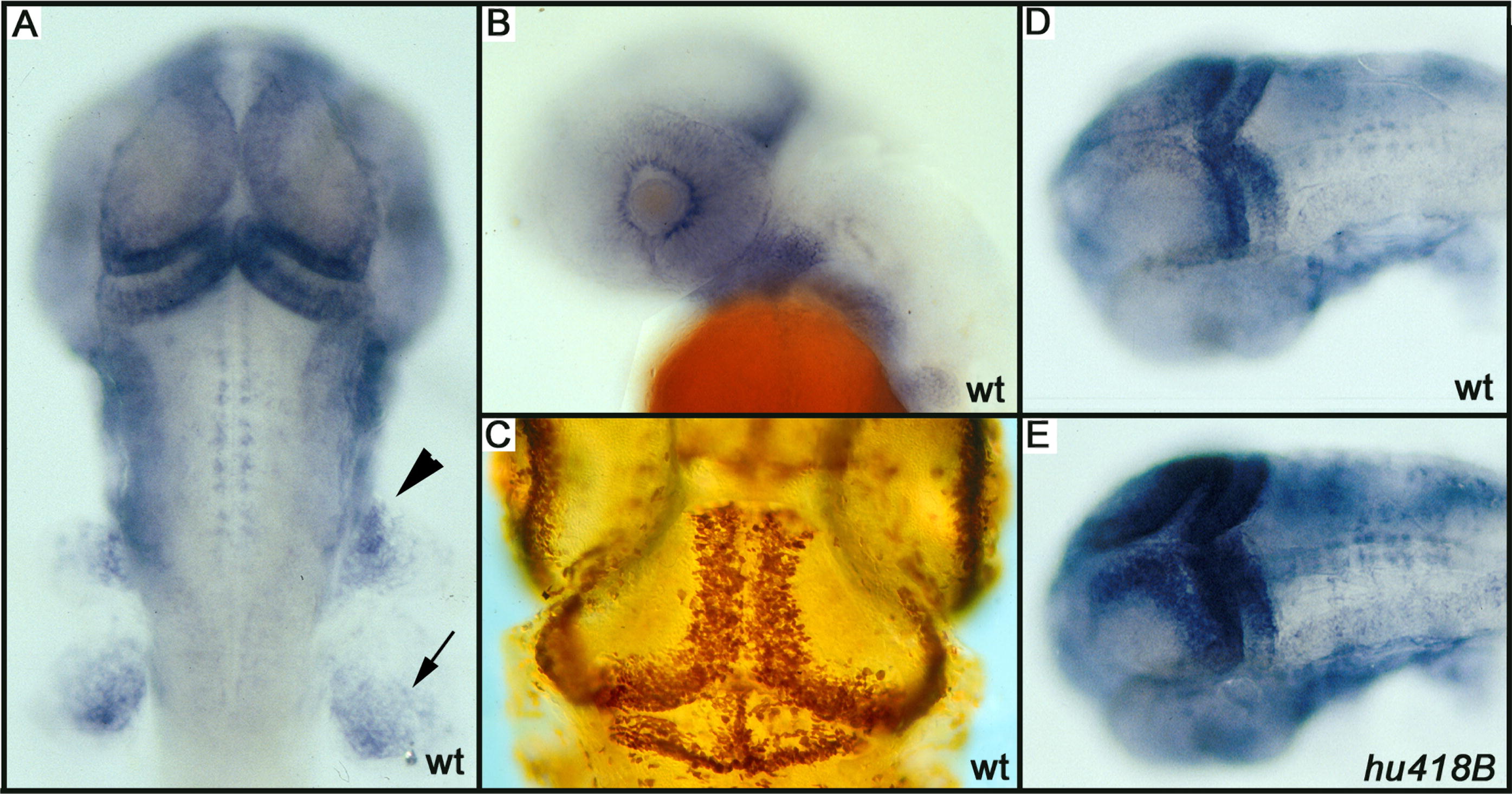
Fig. 1
PCNA and Proliferation Patterns
(A) PCNA pattern as scored during the screen. In a dorsal view at 40 hpf, PCNA staining is observed in the medial and posterior part of the tectum, and in the cerebellum, the neural crest (arrowhead), and the pectoral fin (arrow).
(B) In a sideview (42 hpf), a ring of positive cells is visible around the lens.
(C) In a whole-mount BrdU labeling from day 3.5 to 4.5, similar regions are labeled indicating that PCNA RNA expression prefigures where BrdU will be incorporated.
(D and E) Sibling and hu418B mutants, respectively, showing increased PCNA labeling in the CMZ, but most prominently in the tectum.