
|
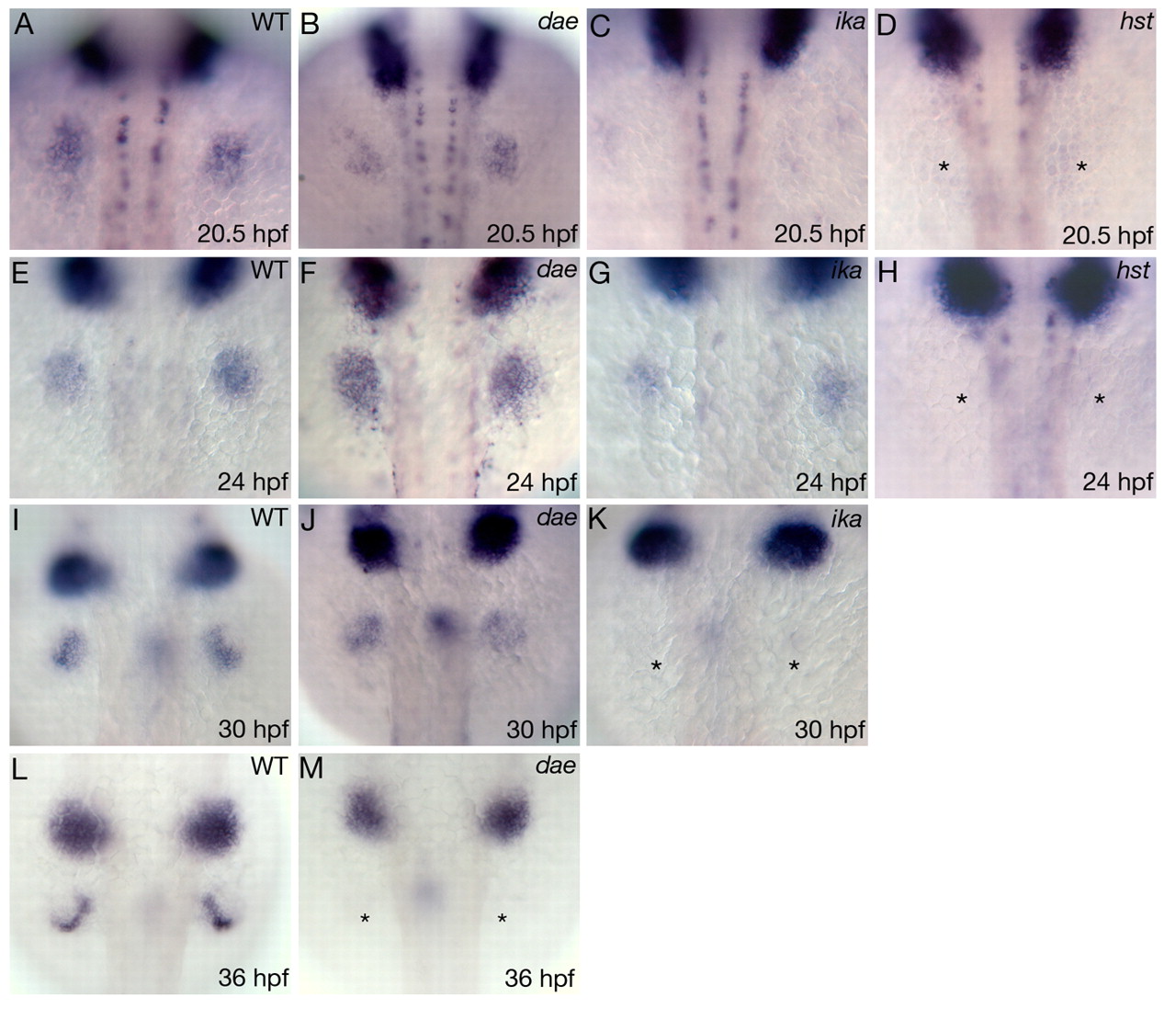
Fig. 6 prdm1 expression in dae, ika and hst mutant embryos. (A-M) Dorsal views of prdm1 expression in pectoral fin buds at 20.5 hpf (A-D), 24 hpf (E-H), 30 hpf (I-K) and 36 hpf (L,M) of wild-type, hst, ika and dae mutants embryos, as indicated in each panel. In dae embryos, activation of prdm1 expression is normal (B,F,J) but its expression disappears at 36 hpf (asterisks, M). In ika, weak prdm1 staining can be detected within the fin field at 24 hpf (G). This expression disappears in 30 hpf embryos (asterisks, K). In hst, prdm1 cannot be detected at any stage analysed (marked by asterisks in D and H). dae, daedalu; hst, heartstrings; i ka, ikarus; WT, wild type.