
|
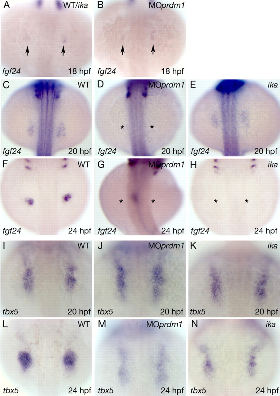
Fig. 5 Expression of fgf24 and tbx5 in MOprdm1-injected embryos compared with ika mutants. (A-H) Dorsal views of fgf24 whole-mount in situ hybridization on wild-type, prdm1 morphant or ikarus mutant embryos, as indicated in each panel. Anterior is to the top. Fgf24 expression is reduced at 18 hpf (B) and is not visible in pectoral fin regions of 20 hpf (D) or 24 hpf (G) MOprdm1-injected embryos. Although the onset of fgf24 expression is normal in fgf24 mutant ikarus embryos (A,E), its expression is not maintained at later stages (H). Asterisks indicate missing fgf24 expression. (I-N) Dorsal views of tbx5 in situ hybridizations on 20 hpf (I-K) and 24 hpf (L-N) wild-type, ika and MOprdm1 embryos. At 20 hpf, no difference can be detected between wild-type, MOprdm1 and ika embryos. At 24 hpf, tbx5 expression is strongly reduced in MOprdm1 and ika fin buds. ika, ikarus; MOprdm1, prdm1 morphant embryo; WT, wild type.