
|
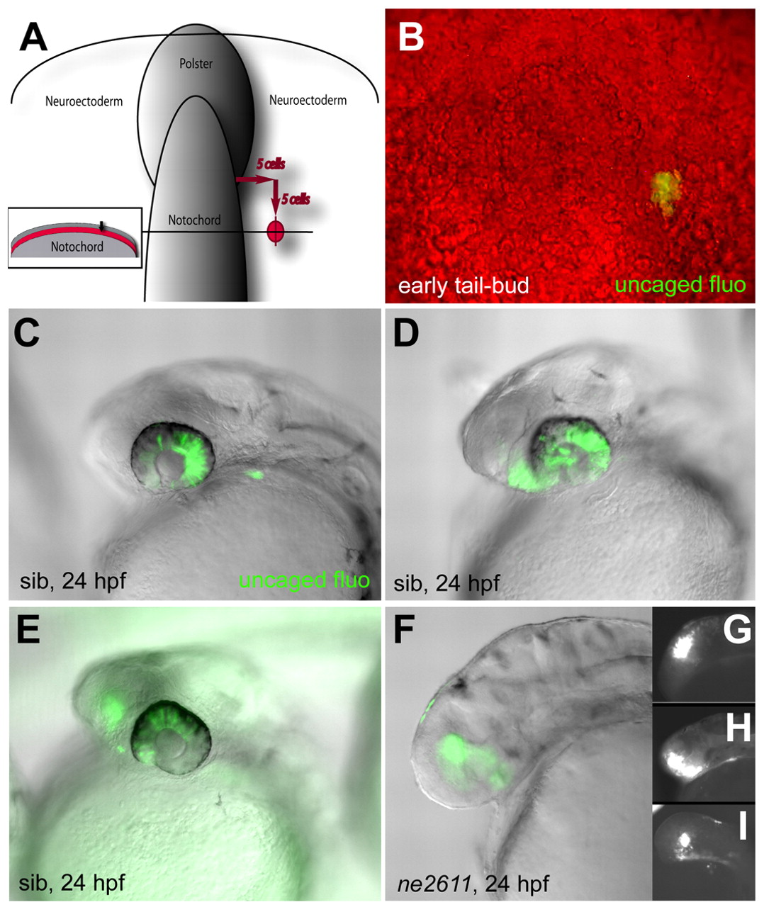
Fig. 6 Cells of the presumptive eye field acquire a telencephalic identity in the absence of Rx3. (A,B) Location of uncaged cells compared with the notochord, polster and enveloping layer (inset in A) at the early tail-bud stage, and photomicrograph of the fluorescent clone immediately following uncaging (B) (dorsal views, anterior up). (C-E) Fate of the uncaged progeny in wild-type embryos at 24 hpf (overlay of fluorescence and Nomarski optics views, anterior left). (C) Retina only (45% of cases); (D) retina and anterior hypothalamus (21% of cases); (E) retina and telencephalon (31% of cases) (see also Table 1). (F-I) Fate of the uncaged progeny in ckhne2611 embryos at the 24 hpf stage (overlay of fluorescence and Nomarski optics views in F, fluorescence only in G-I, anterior left). All clones contribute to the telencephalon (100% of cases, see Table 1).