
Image
|
Figure Caption
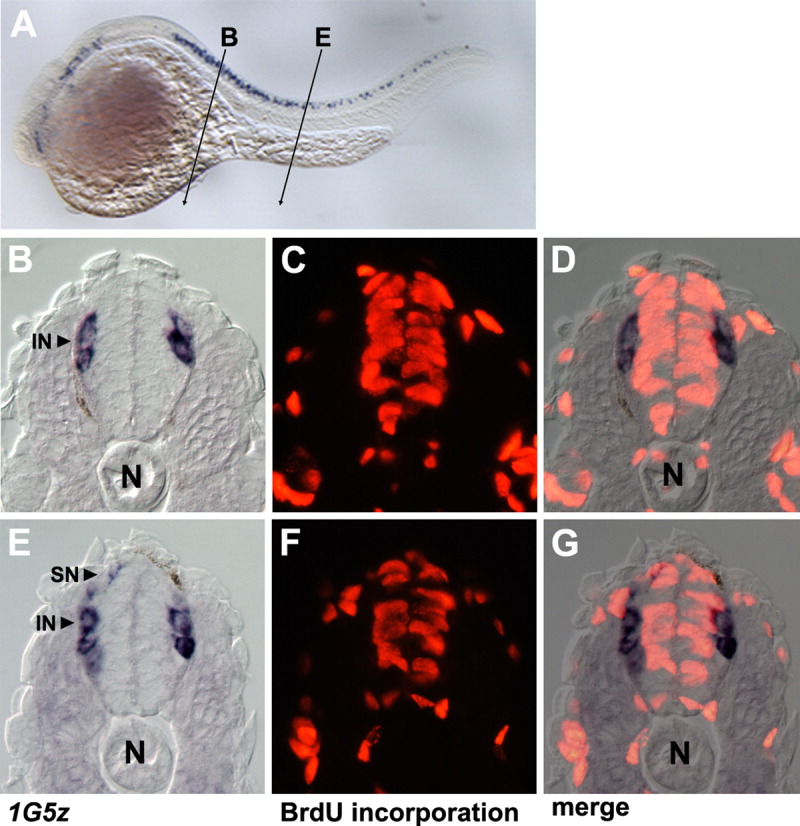
Fig. 5 1G5z-expressing cells are postmitotic neurons. A: Diagram showing the plane of transverse sections (10 μm) in B-D and E-G. B: 1G5z-positive cells in the anterior spinal cord. C: Embryonic tissue in A incubated with bromodeoxyuridine (BrdU). D: Merged image of B and C. Note that 1G5z-stained cells are negative for BrdU staining. E: 1G5z-expressing cells in the medial trunk. F: Embryonic tissue in E treated with BrdU. G: Overlapping image of E and F. Note that 1G5z-positive cells avoid BrdU incorporation. The embryo is fixed at 24 hpf. IN, interneuron; N, notochord; SN, sensory neuron.
Figure Data
Acknowledgments
This image is the copyrighted work of the attributed author or publisher, and
ZFIN has permission only to display this image to its users.
Additional permissions should be obtained from the applicable author or publisher of the image.
Full text @ Dev. Dyn.